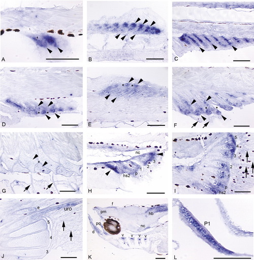
A-L: Expression of sox9a in a developmental fin series (A-J; 4.0 mm NL, 11.5 mm) and head and early pectoral fin (K,L; positive controls).A: Mesenchymal condensations that will give rise to the first two to three anal fin radials express sox9a before cartilage differentiation (4.3 mm NL). B: The stacked chondrocytes of the dorsal fin radials express sox9a (bottom row of arrowheads). Expression is very strong in "balls" of distal mesenchyme (top row arrowheads) that presumably will be incorporated into developing radials (5.3 mm). C: Stacked chondrocytes of the anal fin radials strongly express sox9a, as does distal mesenchyme (not shown in this plane of section, but see B; 5.4 mm). D,E: In both the anal (D) and dorsal (E) fins, as distal radials (D, bottom arrowheads; E, top arrowheads) segment from proximal radials (D, top arrowheads; E, bottom arrowheads), cells in the zone of segmentation (ZS; see *) exhibit down-regulation of sox9a expression. Expression in anal and dorsal fin rays is not detected (fin rays not visible in these planes of section). F: Segmentation is complete. Chondrocytes in distal and proximal radials (arrowheads) continue to express sox9a, while it is not detected in the ZS (*) or in fin rays (arrows; 6.4 mm). G: As radial ossification progresses, expression of sox9a in dorsal and anal fins tapers off in zebrafish > 1 month postfertilization (11.5 mm). H: Below the pre-flexion notochord (nc), hemal spine 2 (hs2), the parhypural (p), and hypurals 1-3 are present and express the gene. Additionally, mesenchyme anterior to hemal spine 2, posterior to hypural 3, and distal to each caudal cartilage condensation strongly expresses sox9a (4.0 mm NL). I: Caudal fin expression is maintained in distal mesenchyme and distal proliferative chondrocytes of the hypurals (1-5), parhypural (p), and hemal spine 2 (hs2), and is observed in proximal chondrocytes of the parhypural and hemal spine 2 (arrowheads). Intermediate regions of the caudal fin supports have begun to ossify (Bird and Mabee,[2003]) and do not express sox9a. Expression in the caudal fin rays is not detected by in situ hybridization (arrows; 5.9 mm). J: In the ossifying caudal fin, expression is no longer detected in hypurals, although it is observed in the developing uroneural (uro) and epural (e; 11.5 mm). K: Expression of sox9a is observed in all cranial cartilages: shown here are the dentary (d), premaxilla (pm), palatoquadrate (pq), and branchial arches (hollow arrowheads). Additional domains of expression are seen in anterior notochord (nc) and in neural cells of the hindbrain (hb) and below the frontals (f) (4.0 mm NL). L: Chondrocytes in early pectoral fin (P1) strongly express sox9a (positive control; Yan et al.,[2002]). Scale bars = 0.1 mm.
|
