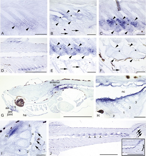
Sagittal sections showing expression of bapx1 in the median fins, head, and trunk of zebrafish. In all panels, anterior is to the left and dorsal to the top. Black patches are melanophores or delimit one of the eyes (G). Arrows mark fin rays, solid arrowheads mark radials, and caret marks mark neural arches. The brown fiber in the top center of C and blue dot in hypural 2 (I) are artifacts. A: In newly forming anal fin radials (5.2 mm), bapx1 expression is observed in the distal chondrocytes and perichondrial cells. B: As radial development progresses (5.6 mm), distal chondrocytes of the proximal radials (top row of arrowheads) continue to express bapx1. bapx1 is not expressed in the ZS (*), the newly formed distal radials (bottom row of arrowheads), or the newly developing fin rays (arrows). C: By 6.4 mm, the distal radials (bottom row of arrowheads) begin to exhibit bapx1 expression, although not at the levels observed in the distal portions of the proximal radials (top row of arrowheads). The ZS (*) does not express the gene. D: The distinct expression pattern of bapx1 is clearly apparent in this section (6.7 mm). Note expression in the distal chondrocytes of the proximal radials, lack of expression in the proximal chondrocytes of the proximal radials and the ZS, and an increase in expression in the distal radials. E: In this 6.8-mm specimen, distal radial bapx1 expression is stronger (bottom row of arrowheads); fin rays do not express the gene (arrows). F: ∼9.0 mm SL: bapx1 expression is no longer detectable in the dorsal and anal fin supports and axial skeleton (9.4 mm). G: At 4.5 mm NL, bapx1 expression in the anterior notochord (nc) is strong. Additional expression domains are observed in the developing posterior articular (aa) and ventral quadrate (q) bones, which together form the jaw joint. Expression is not observed in the anterior anguloarticular or dorsal quadrate, or in the hyoid arch (ha). H: Tissue surrounding the posterior notochord strongly expresses bapx1 (4.5 mm NL). The parhypural (p) and hypurals 1-3 are present, but do not express the gene. I: Posterior notochord expression remains strong at 5.2 mm. All five hypurals are present (2-5 shown); neither hypurals nor caudal fin rays (arrows) express bapx1. J: Neural and hemal arches (∧) and spines (not visible in this plane of section) and the posterior notochord express bapx1 at 6.7 mm. Note continued lack of expression in the fin rays (arrows) and caudal fin supports (curly brackets, inset). Scale bars = 0.5 mm in D,G,J, 0.05 mm in all others.
|
