
Fig. 5
- ID
- ZDB-FIG-070822-48
- Publication
- Kiener et al., 2007 - Identification, tissue distribution and developmental expression of tjp1/zo-1, tjp2/zo-2 and tjp3/zo-3 in the zebrafish, Danio rerio
- Other Figures
- All Figure Page
- Back to All Figure Page
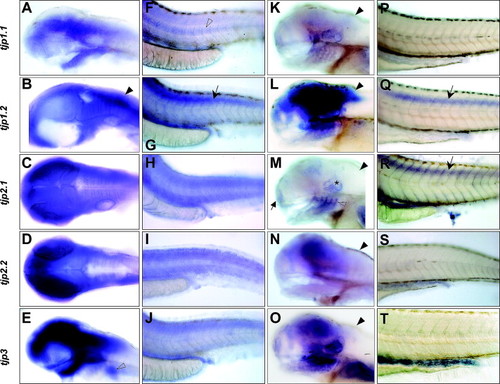
Zebrafish TJP expression pattern at the hatching and larval stages. Expression of tjp1.1 (A,F,K,P), tjp1.2 (B,G,L,Q), tjp2.1 (C,H,M,R), tjp2.2 (D,I,N,S) and tjp3 (E,J,O,T) was detected by in situ hybridization on whole mount embryos at the hatching stage (2 dpf) (A–J) and the larval stage (4 dpf) (K–T). Brain regions express high levels of all TJP genes (A–E). Note that tjp1.2 expression is predominant in the hindbrain (B, arrowhead). The epidermis, the somites and the pronephric ducts also express all five TJPs (F–J). The notochord retains tjp1.1 expression (F, open arrowhead). During the hatching period, the spinal cord starts to express tjp1.2 (G, arrow). The pectoral fin buds are tjp3 positive (E, arrowhead). At the beginning of the larval stage, the expression pattern of the five TJP genes converges. In the anterior part of the embryo, all five TJP genes are now expressed in the nose (arrow), otic capsule (asterisk), branchial arches (open arrowhead), and regions of the brain (K–O, see M). Tjp1.2 is the dominant gene expressed in the brain (L). As already observed at 2 dpf, tjp1.1 and tjp1.2 are expressed in the hindbrain (K,L, arrowheads), from where the other isoforms are absent (M,N,O, arrowheads). In the posterior part of the embryo, the pronephric ducts and intestine highly express TJP genes. Expression in the epidermis and somites is low and not readily detected by in situ hybridization (P–T), but the spinal cord now expresses high levels of both tjp1.2 and tjp2.1 (Q,R, arrows). |
| Genes: | |
|---|---|
| Fish: | |
| Anatomical Terms: | |
| Stage Range: | Long-pec to Day 4 |
Reprinted from Gene expression patterns : GEP, 7(7), Kiener, T.K., Sleptsova-Friedrich, I., and Hunziker, W., Identification, tissue distribution and developmental expression of tjp1/zo-1, tjp2/zo-2 and tjp3/zo-3 in the zebrafish, Danio rerio, 767-776, Copyright (2007) with permission from Elsevier. Full text @ Gene Expr. Patterns