
FIGURE
Fig. 8
Fig. 8
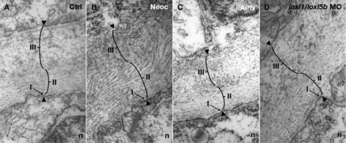
Electron micrographs of the notochord sheath from cross-sections of 30 hpf embryos: (A) 7.4 ng control morpholino; (B) 10 μM neocuproine; (C) 10 mM β-aminopropionitrile; (D) 3.7 ng each of loxl1 and loxl5b morpholino. Sheath components include a basal lamina (I), fibrillar layer (II), and granular layer (III). All three layers are preserved after lysyl oxidase inhibition. |
Expression Data
Expression Detail
Antibody Labeling
Phenotype Data
Phenotype Detail
Acknowledgments
This image is the copyrighted work of the attributed author or publisher, and
ZFIN has permission only to display this image to its users.
Additional permissions should be obtained from the applicable author or publisher of the image.
Reprinted from Developmental Biology, 307(2), Gansner, J.M., Mendelsohn, B.A., Hultman, K.A., Johnson, S.L., and Gitlin, J.D., Essential role of lysyl oxidases in notochord development, 202-213, Copyright (2007) with permission from Elsevier. Full text @ Dev. Biol.