
FIGURE
Fig. S6
- ID
- ZDB-FIG-070802-11
- Publication
- Tessmar-Raible et al., 2007 - Conserved sensory-neurosecretory cell types in annelid and fish forebrain: insights into hypothalamus evolution
- Other Figures
- All Figure Page
- Back to All Figure Page
Fig. S6
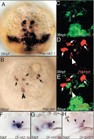
Tryptophane hydroxylase localize to the nk2.1+ region. (A-E) Apical views on the Platynereis forebrain, ventral to the bottom. E is a merged views of C and D. Arrowhead points at Platynereis tryptophan hydroxylase (Pdu-tph) positive (i.e. serotonergic) cells. (F-H) Correlation of serotonergic and nk2.1 cells in the fish hypothalamus. Arrowheads point at tph cell co-expressing nk2.1. Arrows point at cells not co-expressing nk2.1. Riboprobes and stages as indicated. e: epiphysis. |
Expression Data
| Genes: | |
|---|---|
| Fish: | |
| Anatomical Term: | |
| Stage: | Protruding-mouth |
Expression Detail
Antibody Labeling
Phenotype Data
Phenotype Detail
Acknowledgments
This image is the copyrighted work of the attributed author or publisher, and
ZFIN has permission only to display this image to its users.
Additional permissions should be obtained from the applicable author or publisher of the image.
Reprinted from Cell, 129(7), Tessmar-Raible, K., Raible, F., Christodoulou, F., Guy, K., Rembold, M., Hausen, H., and Arendt, D., Conserved sensory-neurosecretory cell types in annelid and fish forebrain: insights into hypothalamus evolution, 1389-1400, Copyright (2007) with permission from Elsevier. Full text @ Cell