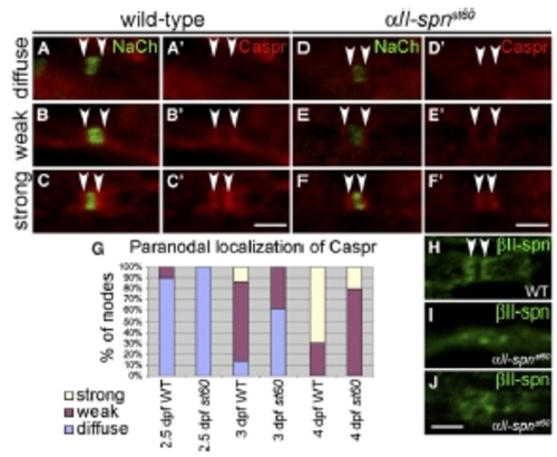
Expression of Paranodal Markers in αII-spectrin Mutants (A–F) PLLn nodes from embryos labeled with anti-panNavCh and anti-Caspr. Anti-Caspr labeling alone is shown in (A′)–(F′). Localization of Caspr to paranodes (indicated by arrowheads) was either diffuse (A and D), weak (B and E) or strong (C and F) in both the wild-type (A–C) and αII-spnst60 mutants (D–F). (G) A histogram showing the proportion of nodes with diffuse, weak, or strong paranodal localization of Caspr at 2.5 dpf (177 nodes from 18 WT embryos and 72 nodes from eight st60 embryos), 3 dpf (102 nodes from eight WT embryos and 105 nodes from five st60 embryos) and 4 dpf (104 nodes from four WT embryos and 98 nodes from four st60 embryos). The accumulation of Caspr at the paranodes in aII-spnst60 mutants lags behind that in the wild-type at each stage. (H–J) PLLn nodes from 5 dpf larvae labeled with anti-βII-spectrin. Anti-βII-spectrin clearly labels paranodes in the wild-type (indicated by arrowheads in [H]), whereas in αII-spnst60 mutants, localization of βII-spectrin is less distinct (I and J). Scale bars represent 2 μm.
|
