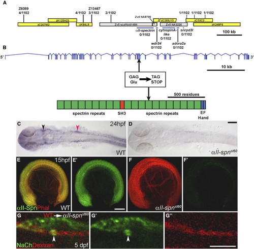
The st60 Mutation Disrupts the αII-spectrin Gene, which Is Required Autonomously in Neurons for Sodium-Channel Clustering (A) Positions of genetic markers along LG21 are shown with the number of recombinations out of 1102 meioses. BACs are indicated by yellow boxes. The st60 mutation maps to a gap in the finished genomic sequence. A rough contig in the gap was built by alignment of sequences from various sources, including genomic scaffold sequence (gray boxes, www.ensembl.org), BACs, and cDNAs. Linkage to st60 was tested for five genes in the gap (indicated by blue bars). (B) The intron-exon structure of αII-spectrin was determined by comparison of cDNA (GenBank accession number EF375552) and genomic sequences. The domain structure of the αII-spectrin polypeptide is also shown. Sequencing of the exons revealed a nonsense mutation in exon 33, which truncates the ORF at amino acid 1491. (C and D) RNA in situ hybridization detecting αII-spn transcripts in the wild-type (C) and αII-spnst60 mutant zebrafish (D) at 24 hpf (dorsal view with anterior left). In the wild-type, expression of αII-spn is widespread and especially prominent in the brain, eyes, and cranial ganglia. Arrowheads mark the region of trigeminal (shown in black) and PLL (shown in red) ganglia. Expression of αII-spn is greatly reduced in αII-spnst60 mutants. The scale bar represents 100 μm. (E–F) Wild-type (E) and αII-spnst60 mutant (F) embryos (anterior left, dorsal up) labeled with phalloidin (phal) and anti-αII-spectrin at 15 hpf. (E2) and (F2) show anti-αII-spectrin labeling alone. Expression of αII-spectrin is ubiquitous in wild-type, whereas expression is nearly abolished in αII-spnst60 mutants. The scale bar represents 100 μm. (G, G2, and G22) Chimeric larvae generated by transplantation of fluorescently labeled wild-type cells into αII-spnst60 mutants were analyzed for the ability to form sodium-channel clusters with the panNavCh antibody. A representative chimera is shown in (G) in which a single wild-type axon from the PLLn is labeled with the lineage marker (red dextran in [G] and [G22]) and has a morphologically normal sodium-channel cluster (indicated by arrowhead, shown in green in [G] and [G2]). The scale bar represents 5 μm.
|
