
Fig. S3
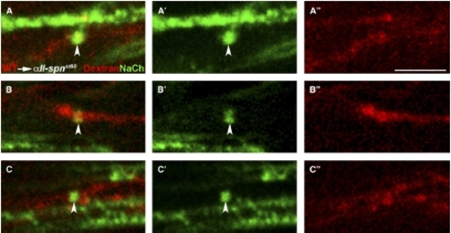
Sodium-Channel Clustering Requires αII-spectrin Function Autonomously in Neurons Chimeric larvae generated by transplantation of fluorescently labeled wild-type cells into αII-spnst60 mutants were analyzed for the ability to form sodium-channel clusters with the panNavCh antibody. Sodium-channel expression (A′–C′), dextran-dye-labeled axons (A″–C″), and merged images (A–C) are shown from the PLLn of three different chimeras. In these examples, all of the dye-labeled cells are neurons and normal sodium-channel clusters (indicated by arrowheads) are only evident in dye-labeled wild-type axons. Unmyelinated axons, which have sodium-channel proteins throughout the axolemma, can also be seen (e.g., in [A]). The genotype of mutant hosts was confirmed by PCR. The scale bar represents 5 μm. |