
Fig. 2
- ID
- ZDB-FIG-060630-1
- Publication
- Gillhouse et al., 2004 - Two Frodo/Dapper homologs are expressed in the developing brain and mesoderm of zebrafish
- Other Figures
- All Figure Page
- Back to All Figure Page
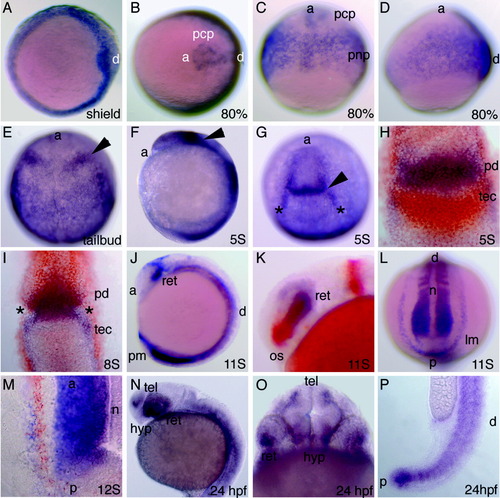
Figure 2. Expression of frd1 during gastrulation and somitogenesis. Staged embryos were stained for frd1 (purple), mbx (orange in H), dlx3 (orange in I), and pax2 (orange in K) RNA using whole-mount in situ hybridization. A: Early gastrula (shield) stage embryos, animal pole view, dorsal to the right. Frd1 is expressed around the circumference of the blastoderm margin and in the forming dorsal shield. B-D: Mid-gastrula (80% epiboly) stage embryos: B, animal pole view, dorsal to the right; C, dorsal view, anterior at the top; D, lateral view, dorsal to the right. frd1 is expressed in the posterior dorsal neurectoderm and in the axial mesoderm, including the prechordal plate (pcp). E: End of gastrulation (tail bud) stage embryo, dorsal view. Frd1 is found in a new domain of expression corresponding to the future posterior diencephalon (arrowhead). Strong expression is also seen throughout the trunk. F-I: Early somitogenesis (five- to eight-somite) stage embryos, dorsal view, anterior at the top. F: Lateral view, anterior at the top. Expression continues in the future posterior diencephalon (arrowhead). G: Anteriorly, strong frd1 expression is seen in a stripe in the posterior dorsal diencephalon and in two stripes at the lateral borders of the anterior brain (asterisks). H: The stripe of frd1 expression in the posterior diencephalon (pd) is located just anterior to the tectal domain of mbx expression (tec). I: Lateral expression domains of frd1 (asterisks) are found just medial to dlx3 expression, in a position corresponding to presumptive head mesenchyme. J-M: Mid-somitogenesis (11- to 12-somite) stage embryos. J,K: Lateral view, anterior to the left, dorsal at the top. J:frd1 is expressed strongly in the forming retina (ret) and posterior mesoderm (pm) and weakly in the trunk mesoderm. K: The retinal expression domain of frd1 (ret) is found just posterior to the optic stalk marked by expression of pax2 in orange (os). L,M: Posterior dorsal view, dorsal at the top. L:frd1 is expressed in paraxial mesoderm (somites and segmental plate) and in a stripe of expression corresponding to lateral mesoderm (lm). Note absence of expression in the axial mesoderm (n). M: Lateral mesoderm expression of frd1 is found lateral to pax2 (orange) expression in intermediate mesoderm, the forming pronephric ducts. N-P: Late somitogenesis (24 hr postfertilization [hpf]) embryos. N: Lateral view, dorsal at the top. O: Anterior view, dorsal at the top. N,O:Frd1 is expressed in distinct domains in the telencephalon (tel), hypothalamus (hyp), and ventral retina (ret). P: Lateral view of tail bud expression, anterior at the top. a, anterior; d, dorsal; hyp, hypothalamus; n, notochord; p, posterior; pnp, posterior neural plate. Asterisk indicates head mesenchyme expression. |