
FIGURE
Fig. 1
- ID
- ZDB-FIG-051209-2
- Publication
- Prabhudesai et al., 2005 - Targeted effects of retinoic acid signaling upon photoreceptor development in zebrafish
- Other Figures
- All Figure Page
- Back to All Figure Page
Fig. 1
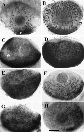
Effects of RA on rod and cone opsin expression. Panels show lateral views of whole-mounted eyes from DMSO-treated zebrafish embryos (A, C, E, G) or embryos treated with RA (B, D, F, H) at 51 hpf and fixed at 75 hpf. Embryos were hybridized with rod opsin riboprobes (A and B), red cone opsin riboprobes (C and D), blue cone opsin riboprobes (E and F), or UV cone opsin riboprobes (G and H). v, ventral (for all panels); scale bar = 40 μm; * indicates slight damage to eye tissue during processing. |
Expression Data
| Genes: | |
|---|---|
| Fish: | |
| Condition: | |
| Anatomical Terms: | |
| Stage: | Protruding-mouth |
Expression Detail
Antibody Labeling
Phenotype Data
Phenotype Detail
Acknowledgments
This image is the copyrighted work of the attributed author or publisher, and
ZFIN has permission only to display this image to its users.
Additional permissions should be obtained from the applicable author or publisher of the image.
Reprinted from Developmental Biology, 287(1), Prabhudesai, S.N., Cameron, D.A., and Stenkamp, D.L., Targeted effects of retinoic acid signaling upon photoreceptor development in zebrafish, 157-167, Copyright (2005) with permission from Elsevier. Full text @ Dev. Biol.