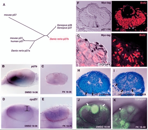
Canonical Wnt signalling is required for PKA-mediated cell proliferation. (A) Phylogenetic relationship among vertebrate p27 proteins and mouse p57. The length of each branch is proportional to sequence divergence from the branch points. (B,C) In situ hybridisation of 36-hpf DMSO- and forskolin-treated retinas with a p27b RNA probe. p27b is expressed in the DMSO-treated retina (B) but not in the forskolin-treated retina (C). (D,E) In situ hybridisation of 36-hpf DMSO-treated (D) and forskolin-treated (E) retinas with a cyclin D1 RNA probe. cyclin D1 is downregulated in differentiating neurons and is localised in the CMZ of the DMSO-treated retina (D, lines/arrows). However, cyclin D1 is not downregulated in the forskolin-treated retina (E) but remains expressed in a large region. (F) 48-hpf forskolin-treated embryos expressing myc-tagged p27 labelled with the anti-myc antibody (brown in left panel) and anti-BrdU antibody (red in right panel). Retinal cells expressing p27 are BrdU negative (white asterisks), whereas almost all p27-negative cells incorporate BrdU. (G) 33-hpf forskolin-treated embryos expressing myc-tagged N-Tcf3 labelled with the anti-myc antibody (brown in left panel) and anti-BrdU antibody (red). Retinal cells expressing ΔN-Tcf3 are BrdU negative (white asterisks). (H,I) 48-hpf wild-type retinas expressing Δ47-ß-catenin (H) and a mixture of Δ47-ß-catenin and dnPKA (I). Multi-folded neural retina is observed (arrows) in both cases, suggesting that dnPKA does not inhibit Wnt-induced proliferation. (J,K) GFP expression in 2-dpf TOPdGFP transgenic retinas treated with DMSO (J) and forskolin (K). GFP is expressed in the CMZ of the DMSO-treated retina (J, arrows and white dashed lines). In the forskolin-treated retina, GFP expression is not detected. Arrowhead (K) indicates GFP expression in epidermis.
|
