
Fig. 5
- ID
- ZDB-FIG-050518-11
- Publication
- Masai et al., 2005 - The hedgehog-PKA pathway regulates two distinct steps of the differentiation of retinal ganglion cells: the cell-cycle exit of retinoblasts and their neuronal maturation
- Other Figures
- All Figure Page
- Back to All Figure Page
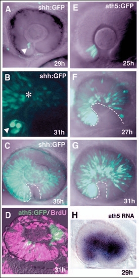
Pattern of progression of shh:GFP and ath5:GFP expression in zebrafish retina. (A-C) Lateral view of the optic cup of the transgenic line Tg(shh:GFP). B is shown at twice the magnification of A and C. shh:GFP is expressed in a few cells adjacent to the optic stalk at 29 hpf (A, white arrowhead). Progression of GFP expression is observed in the adjacent dorsal region at 31 hpf (B, asterisk). A wave front of shh:GFP expression reaches the temporal region of the neural retina at 35 hpf (C, dotted line). (D) Double labelling of 31-hpf retinas of the transgenic line Tg(ath5:GFP) with the anti-BrdU antibody and ath5:GFP antibody. ath5:GFP-positive cells (green) are BrdU (magenta) negative. (E-G) GFP expression in the transgenic line Tg(ath5:GFP). Throughout the stages examined, the pattern of ath5:GFP expression is similar to that of ath5 transcription. Note that GFP-expressing cells are already observed in the temporal region at 27 hpf (F), and the density of GFP-positive cells increases in later stages (G). The dotted line indicates a wave front of ath5:GFP expression. (H) ath5 RNA expression in the transgenic line Tg(shh:GFP) at 29 hpf. This is the same embryo that is shown in A. ath5 expression spreads to the large region of the neural retina. |