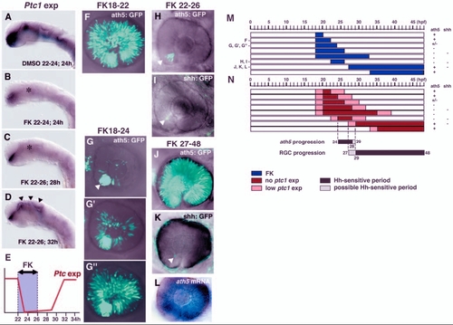
Hh signalling regulates two distinct steps of RGC differentiation. (A,B) In situ hybridization of 24-hpf embryos with a ptc1 RNA probe. Embryos were treated with DMSO (A) and forskolin (B) from 22 to 24 hpf. ptc1 expression is observed in the ventral CNS in DMSO-treated embryos (A), but is drastically reduced in forskolin-treated embryos (B, asterisk), although weak expression is still observed in the ventral forebrain. (C,D) ptc1 expression of embryos treated with forskolin between 22 and 26 hpf. ptc1 expression is still low at 28 hpf (C, asterisk), but has recovered at 32 hpf (D, arrowheads). (E) Schematic diagram of the time lag between forskolin treatment and inhibition of the Hh signalling pathway. ptc1 expression is inhibited within 2 hours of the start of forskolin treatment. ptc1 expression is still low 2 hours after the removal of forskolin, but has recovered within 6 hours of the removal of forskolin. (F) ath5:GFP expression in retina treated with forskolin from 18 to 22 hpf; ath5:GFP expression progresses throughout the neural retina. (G-G′′) ath5:GFP expression in retinas treated with forskolin from 18 to 24 hpf. The inhibition of progression varies from severe (G), to mild (G′) and nearly normal (G′′). In the severe case, ath5:GFP is observed only in the ventronasal retina (white arrowhead). (H) ath5:GFP expression in retina treated with forskolin from 22 to 26 hpf. The progression of ath5:GFP expression is completely blocked (white arrowhead). (I) shh:GFP expression in retina treated with forskolin from 22 to 26 hpf. shh:GFP expression also fails to progress from the ventronasal retina (white arrowhead). (J) ath5:GFP expression in retina treated with forskolin from 27 to 48 hpf. The propagation of ath5:GFP expression occurs normally throughout the neural retina. (K) shh:GFP expression in retina treated with forskolin from 27 to 48 hpf. The progression of shh:GFP expression remains blocked (white arrowhead). Weak background signals are observed in the skin surrounding the eye. (L) ath5 RNA expression in the same embryo as shown in K. ath5 RNA expression spreads to whole region of the neural retina. (M) Pulse treatment with forskolin in different time windows. Blue bars indicate the period of treatment with forskolin. All embryos were examined at 48 hpf. +, progression of ath5:GFP or shh:GFP expression occurs normally; – indicates that progression is effectively blocked. (N) Time-window of Hh signalling inhibition, estimated by considering the time lag between forskolin treatment and the downregulation of ptc1 expression. Red bars indicate the period when ptc1 expression is severely suppressed by forskolin treatment. Pink bars indicate the time lag between the start of forskolin treatment and the suppression of ptc1 expression, or between the removal of forskolin and the recovery of ptc1 expression; within these time periods Hh signalling may be reduced. Black and grey bars indicate Hh-sensitive and possible Hh-sensitive periods for the progression of ath5 expression and RGC maturation, respectively. FK, forskolin.
|
