
Fig. 2
- ID
- ZDB-FIG-050214-8
- Publication
- Galloway et al., 2005 - Loss of gata1 but not gata2 converts erythropoiesis to myelopoiesis in zebrafish embryos
- Other Figures
- All Figure Page
- Back to All Figure Page
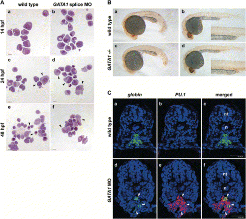
Erythroid Precursors Are Converted into Myeloid Precursors in the Absence of Gata1(A) gata1-gfp cells from wild-type (a) and gata1 splice MO-injected (b) embryos at 10 somites (14 hpf) resemble hematopoietic precursors. At 24 hpf, most wild-type gata1-gfp cells resemble proerythroblasts (c, arrowheads), but myeloid cells with indented nuclei are observed. Cells from gata1 splice MO-injected embryos have myeloid features such as vacuoles and indented nuclei (d, arrowheads). Many erythrocytes and proerythroblasts are isolated from 48 hpf wild-type embryos (e, arrowheads) as well as a few myeloid cells. Cells from 48 hpf gata1 splice MO-injected embryos have vacuoles and indented nuclei (f, arrowheads), suggesting that they are myelomonocytes (scale bars are 10 μm).(B) Wild-type (a) and vlt mutant (c) embryos at 22 hpf have similar numbers of TUNEL-positive cells. vlt mutant embryos have an approximate 2-fold increase in apoptosis in their ICM region (d, inset) compared to their wild-type siblings (b, inset) at 28 hpf.(C) Confocal imaging of transverse sections of DAPI-stained (blue) embryos that have undergone double in situ hybridization for βe1 globin (green) and pu.1 (red). Wild-type embryos express βe1 globin and not pu.1 in their ICM cells at 22 hpf (a–c). gata1 MO-injected embryos express both βe1 globin and pu.1 in their ICM cells at 22 hpf (d–f). Some cells coexpress (yellow) both genes (d–f, arrowheads; n, notochord; nt, neural tube; scale bar equals 40 μm). |
| Genes: | |
|---|---|
| Fish: | |
| Knockdown Reagent: | |
| Anatomical Term: | |
| Stage: | 26+ somites |
Reprinted from Developmental Cell, 8(1), Galloway, J.L., Wingert, R.A., Thisse, C., Thisse, B., and Zon, L.I., Loss of gata1 but not gata2 converts erythropoiesis to myelopoiesis in zebrafish embryos, 109-116, Copyright (2005) with permission from Elsevier. Full text @ Dev. Cell