
Zebrafish Anatomical Dictionary
Structure description: otolith
by Tanya Whitfield
Name: otolith
Abbreviation: None
Synonyms: None
Figures:
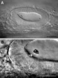 otoliths
Description: Stony accretions of crystalline calcium carbonate and protein. These develop over the anterior and posterior maculae in the ear and are tethered in place by a gelatinous otolithic membrane, into which the macular hair cells project. From 18-22h, many small crystalline particles are present throughout the lumen of the ear, where they are agitated and distributed by beating cilia. From 18.5h onwards, the particles coalesce to form the two otoliths; these initially appear as irregular clumps of material at the anterior and posterior ends of the otic vesicle. After 24h, few free particles are observed, and the otoliths grow in size, acquiring distinctive shapes and sizes during the larval period.
Homologues:
- Human: otoconia
- Mouse: otoconia
- Chicken: otoconia
- Frog: otoliths
- Fly: none
Stages:
- First appears at: 18h
- Disappears (or changes name) at: Unknown
Parents (forms from): Supporting cells are thought to give rise to the correct milieu in which the otoliths can precipitate. Hair cells or non-sensory cell, may also play a role (see Haddon et al., 1999). Otoliths nucleate over hair cell precursors ("tether cells") at the anterior and posterior ends of the otic vesicle (Riley et al., 1997).
Children:
- Presumptive (thought to give rise to): Adult otoliths. The adult fish has 3 otoliths in each ear: the sagitta of the saccule, the lapillus of the utricle and the astericus of the lagena. The anterior otolith of the embryo is probably the precursor of the lapillus of the adult, and the posterior otolith is probably the precursor of the sagitta. It is not known when the astericus (and its associated sensory patch, the lagena) arise.
- Anlage (known to give rise to): Unknown
Group (member of):
- Anatomical (group member): ear
- Functional (group member): auditory, vestibular system
Markers:
- mRNA: None
- Antibodies: None
- Other: Otoliths are easily visible in the live embryo under dissecting and compound microscopes without the need for a special stain. The free particles at early stages can be visualised in the live embryo with DIC optics using a high magnification objective.
Publications:
- Primary: Haddon, C., and Lewis, J. (1996). Early ear development in the embryo of the zebrafish, Danio rerio. Journal of Comparative Neurology 365, 113-123.
- Secondary:
Haddon, C., Mowbray, C., Whitfield, T., Jones, D., Gschmeissner, S., and Lewis, J. (1999). Hair cells without supporting cells: further studies in the ear of the zebrafish mind bomb mutant. Journal of Neurocytology 28, 837-850.
Comments: None