
- Title
-
The function of silberblick in the positioning of the eye anlage in the zebrafish embryo
- Authors
- Heisenberg, C.P. and Nüsslein-Volhard, C.
- Source
- Full text @ Dev. Biol.
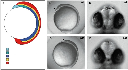
slb embryos are shorter at bud stage. The eyes are turned inward anteriorly at later developmental stages. (A) Schematic drawing of the embryonic body axis organization at bud stage. (B, D) Side view of a wild-type (B) and slb (D) embryo at bud stage. (C, E) Frontal view of a wild-type (C) and slb (E) embryo at 48 hpf. Arrowheads in B and D indicate position of the polster. Anterior to the left. |
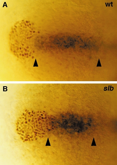
The posterior prechordal plate of slb embryos is shortened and broadened, whereas the polster appears narrower and more elongated at bud stage. (A, B) Double labeling showing cells of the posterior prechordal plate expressing rtk2 (blue, in situ staining) and polster cells stained for Fkd2 (brown, antibody staining) in a wild-type (A) and slb (B) embryo at bud stage. Arrowheads demarcate the length of the posterior prechordal plate along the anterior-posterior axis. Dorsal views, anterior to the left. PHENOTYPE:
|
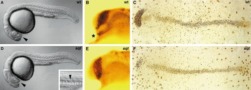
In sqt embryos, the eyes are fused anteriorly and the ventral diencephalon as well as the underlying prechordal plate is reduced. (A, D) Side view of a wild-type (A) and sqt (D) embryo at 24 hpf. (B, E) The axonal scaffold stained with an anti-acetylated tubulin antibody in wild-type (B) and sqt (E) embryos at 24 hpf. (C, F) The axial mesendoderm stained with an anti-Fkd2 antibody in wild-type (C) and sqt (F) embryos at bud stage. Arrowheads in A and D point to the position of the optic stalks. Small picture in the bottom right corner of D shows the presence of a floorplate (arrowhead) in sqt mutants at 24 hpf. Asterisk in B demarcates the position of the nucleus of the tract of the postoptic commissur. Side (A, B, D, E) and dorsal (C, F) views, anterior to the left. |

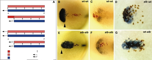
The notochord is affected independently from the polster in slb at bud stage. (A, B) Notochord cells expressing ntl in a sqt mutant (which lacks polster cells, as revealed by probing the embryos for hgg1 expression) (A) and a slb/sqt double mutant (B) at bud stage. Dorsal views, anterior to the left. PHENOTYPE:
|

Posterior displacement of the polster in slb is accompanied by a shortening of the shh expression domain at bud stage. (A, C) Double labeling staining the polster with an anti-Fkd2 antibody (brown) and the anterior edge of the neural plate by its expression of dlx3 (blue) in a wild-type (A) and slb (C) embryo at bud stage. (B, D) In situ staining which labels the midline (shh) and anterior edge of the neural plate (dlx3) in a wild-type (B) and slb (D) embryo at bud stage. Arrowheads in D demarcate the gap between the anterior edge of the neural plate and the anterior end of the shh expression domain in slb. Dorsal views, anterior to the left. PHENOTYPE:
|
Anterior migration of ventral CNS midline cells is normal at bud stage but impaired at 15 hpf in slb. (A) Schematic diagram showing two different models which might explain the shortening of the ventral CNS midline in slb: (I) reduced anterior migration of the axial mesendoderm (blue) in slb leads to incomplete induction of the ventral CNS midline (hatched, red) in the overlying neural plate (red). (II) Anterior migration of both the axial mesendoderm (blue) and the overlying ventral CNS midline (hatched, red) is impaired in slb (B, E) Position of ventral diencephalic midline cells (brown) in relation to the polster expressing hgg1 (blue) in a wild-type (B) and slb (E) embryo at bud stage. Note that the anterior end of the neural keel (arrowhead) lies to the posterior of the polster in wild type whereas it lies to the anterior of the polster in slb. (C, F) Position of ventral diencephalic midline cells (brown) in relation to the anlage of the optic stalks expressing pax2 (blue) in a wild-type (C) and slb (F) embryo at 15 hpf. (D, G) Transplantation of slb cells (brown) into a wildtype polster expressing hgg1 (blue) (D) and of wild-type cells (brown) into a slb polster expressing hgg1 (blue) (G) do neither phenocopy nor rescue the mutant phenotype. Dorsal views, anterior to the left. |
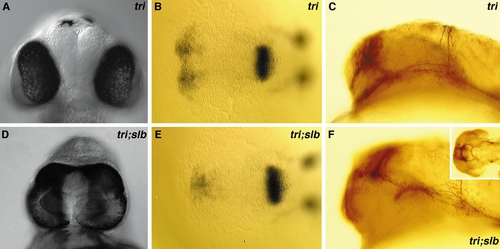
In slb/tri double mutants the slb eye phenotype is strongly enhanced. (A, D) Frontal view of a tri (A) and slb/tri (D) double mutant at 48 hpf. (B, E) The anlage of the optic stalks expressing pax2 in a tri (B) and slb/tri (E) double mutant at 15 hpf. Dorsal views, anterior to the left. (C, F) The axonal scaffold stained for acetylated tubulin appears normal in a tri (C) mutant and is still intact but strongly deformed in a slb/tri (F) double mutant at 24 hpf (the small picture in the upper right corner of F shows a dorsal view of the axonal scaffold stained for acetylated tubulin in slb/tri embryos at 24 hpf). Side views, anterior to the left. PHENOTYPE:
|
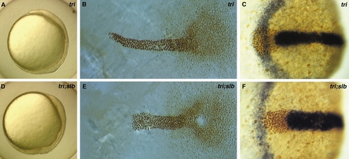
In slb/tri double mutants the slb notochord defect is strongly enhanced, whereas the shape of the polster and the distance between the anterior edge of the neural plate and the anterior end of the shh expression domain is unchanged. (A, D) Live phenotypes of a tri (A) and slb/tri (D) double mutant at bud stage. Side views, anterior to the left. (B, E) The notochord stained for Ntl in a tri (B) and slb/tri (E) double mutant. (C, F) The polster stained for Fkd2 (brown, antibody staining), the ventral CNS midline expressing shh (blue, in situ staining), and the anterior edge of the neural plate expressing dlx3 (blue, in situ staining) in a tri (C) and slb/tri (F) double mutant. Dorsal views, anterior to the left. PHENOTYPE:
|
Reprinted from Developmental Biology, 184(1), Heisenberg, C.P. and Nüsslein-Volhard, C., The function of silberblick in the positioning of the eye anlage in the zebrafish embryo, 85-94, Copyright (1997) with permission from Elsevier. Full text @ Dev. Biol.