
Image
|
Figure Caption
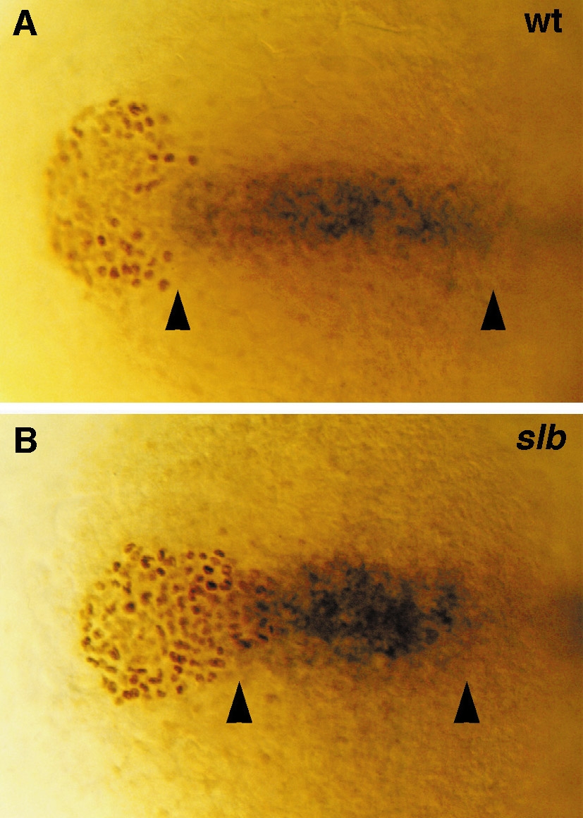
Fig. 2 The posterior prechordal plate of slb embryos is shortened and broadened, whereas the polster appears narrower and more elongated at bud stage. (A, B) Double labeling showing cells of the posterior prechordal plate expressing rtk2 (blue, in situ staining) and polster cells stained for Fkd2 (brown, antibody staining) in a wild-type (A) and slb (B) embryo at bud stage. Arrowheads demarcate the length of the posterior prechordal plate along the anterior-posterior axis. Dorsal views, anterior to the left.
Figure Data
Acknowledgments
This image is the copyrighted work of the attributed author or publisher, and
ZFIN has permission only to display this image to its users.
Additional permissions should be obtained from the applicable author or publisher of the image.
Reprinted from Developmental Biology, 184(1), Heisenberg, C.P. and Nüsslein-Volhard, C., The function of silberblick in the positioning of the eye anlage in the zebrafish embryo, 85-94, Copyright (1997) with permission from Elsevier. Full text @ Dev. Biol.