
Fig. 7
- ID
- ZDB-FIG-090504-59
- Publication
- Heisenberg et al., 1997 - The function of silberblick in the positioning of the eye anlage in the zebrafish embryo
- Other Figures
- All Figure Page
- Back to All Figure Page
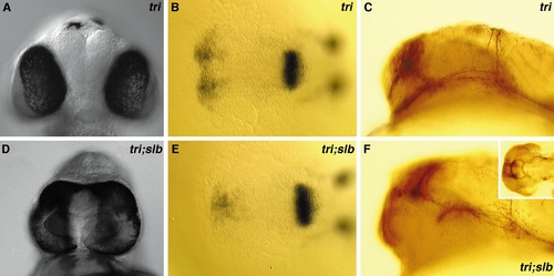
In slb/tri double mutants the slb eye phenotype is strongly enhanced. (A, D) Frontal view of a tri (A) and slb/tri (D) double mutant at 48 hpf. (B, E) The anlage of the optic stalks expressing pax2 in a tri (B) and slb/tri (E) double mutant at 15 hpf. Dorsal views, anterior to the left. (C, F) The axonal scaffold stained for acetylated tubulin appears normal in a tri (C) mutant and is still intact but strongly deformed in a slb/tri (F) double mutant at 24 hpf (the small picture in the upper right corner of F shows a dorsal view of the axonal scaffold stained for acetylated tubulin in slb/tri embryos at 24 hpf). Side views, anterior to the left. |
| Fish: | |
|---|---|
| Observed In: | |
| Stage Range: | 10-13 somites to Long-pec |
Reprinted from Developmental Biology, 184(1), Heisenberg, C.P. and Nüsslein-Volhard, C., The function of silberblick in the positioning of the eye anlage in the zebrafish embryo, 85-94, Copyright (1997) with permission from Elsevier. Full text @ Dev. Biol.