FIGURE SUMMARY
Title

Infection-experienced HSPCs protect against infections by generating neutrophils with enhanced mitochondrial bactericidal activity

Authors
Darroch, H., Keerthisinghe, P., Sung, Y.J., Rolland, L., Prankerd-Gough, A., Crosier, P.S., Astin, J.W., Hall, C.J.
Source
Full text @ Sci Adv

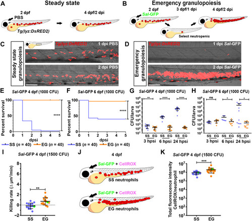
EG larvae have elevated survival to subsequent bacterial challenge and have neutrophils with enhanced bactericidal activity.

(A and B) Schematic illustrating strategy to generate SS (A) and EG (B) larvae. (C and D) Live imaging of the AGM/CHT regions within Tg(lyz:DsRED2) larvae at 1 and 2 dpi with PBS (C) or Sal-GFP (D). (E and F) Kaplan-Meier graphs showing survival of SS and EG larvae over 5 dpsi with Sal-GFP at 4 dpf (E) and 6 dpf (F). (G and H) Bacterial burdens within individual SS or EG larvae at 3, 6, and 24 hpsi with Sal-GFP at 4 dpf (G) and 6 dpf (H). (I) Bacterial killing rates of SS and EG neutrophils following Sal-GFP infection. Green data points highlight killing rates of neutrophils as shown in fig. S1D. (J) Schematic illustrating injection of SS and EG larvae with Sal-GFP and CellROX. (K) Quantification of ROS production within Sal-GFP-laden SS and EG neutrophils, as detected by CellROX fluorescence. Green data points highlight ROS production of neutrophils as shown in fig. S1G. Error bars, mean ± SD; ns, not significant, *P < 0.05, **P < 0.01, ***P < 0.001, ****P < 0.0001; Gehan-Breslow-Wilcoxon test (E and F) and unpaired Student’s t test (G, H, I, and K). Scale bars, 50 μm.

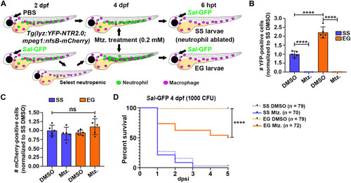
Ablating EG neutrophils reduces survival to subsequent infection.

(A) Schematic illustrating strategy to ablate SS and EG neutrophils. Tg(lyz:YFP-NTR2.0;mpeg1:nfsB-mCherry) SS larvae and those demonstrating EG were treated with dimethyl sulfoxide (DMSO; control) or 0.2 mM mtz. At 6 hours posttreatment (hpt), neutrophil-ablated larvae were infected with Sal-GFP and treated continuously with 0.1 mM mtz. (B) Flow quantification of YFP-expressing neutrophils from whole SS and EG Tg(lyz:YFP-NTR2.0;mpeg1:nfsB-mCherry) larvae, 6 hours following mtz treatment, compared to DMSO-treated controls (n = 20 larvae per sample, five experimental replicates). (C) Flow quantification of mCherry-expressing macrophages from whole SS and EG Tg(lyz:YFP-NTR2.0;mpeg1:nfsB-mCherry) larvae, 6 hours following mtz treatment, compared to DMSO-treated controls (n = 20 larvae per sample, five experimental replicates). (D) Kaplan-Meier graph showing survival of DMSO- and mtz-treated SS and EG Tg(lyz:YFP-NTR2.0;mpeg1:nfsB-mCherry) larvae over 5 dpsi with Sal-GFP at 4 dpf. Error bars, mean ± SD; ****P < 0.0001; one-way analysis of variance (ANOVA) with Tukey’s multiple comparisons test (B and C) and Gehan-Breslow-Wilcoxon test (D).

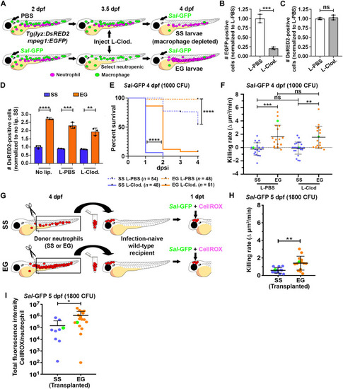
EG neutrophils maintain elevated killing rates in macrophage-depleted larvae and when transplanted into infection-naïve recipients.

(A) Schematic illustrating strategy to deplete macrophages within SS and EG larvae. (B and C) Flow quantification of EGFP-expressing macrophages (B) and DsRED2-expressing neutrophils (C) from whole 4 dpf Tg(lyz:DsRED2;mpeg1:EGFP) larvae, following liposomal-clodronate (L-Clod) injection, compared to liposomal-PBS (L-PBS) injection controls (n = 20 larvae per sample, three experimental replicates). (D) Flow quantification of DsRED2-expressing neutrophils from whole 4 dpf SS and EG Tg(lyz:DsRED2;mpeg1:EGFP) larvae, 1 day following L-Clod. injection, compared to L-PBS injection and no liposome (no lip.) controls (n = 20 larvae per sample, three experimental replicates). (E) Kaplan-Meier graph showing survival of SS L-PBS, SS L-Clod., EG L-PBS, and EG L-Clod. larvae over 4 dpsi with Sal-GFP at 4 dpf. (F) Bacterial killing rates of SS and EG neutrophils within L-Clod.– and L-PBS–injected Tg(lyz:DsRED2;mpeg1:EGFP) larvae following Sal-GFP infection. Green data points highlight killing rates of neutrophils as shown in fig. S4C. (G) Schematic illustrating transplantation of FACS-isolated SS and EG neutrophils into infection-naïve recipient larvae. (H) Bacterial killing rates of transplanted SS and EG neutrophils following Sal-GFP infection. Green data points highlight killing rates of neutrophils as shown in fig. S4D. (I) Quantification of ROS production within Sal-GFP-laden transplanted SS and EG neutrophils, as detected by CellROX fluorescence. Green data points highlight ROS production of neutrophils as shown in fig. S4E. Error bars, mean ± SD; *P < 0.05, **P < 0.01, ***P < 0.001, ****P < 0.0001; unpaired Student’s t test (B, C, D, H, and I), Gehan-Breslow-Wilcoxon test (E), and one-way ANOVA with Tukey’s multiple comparisons test (F).

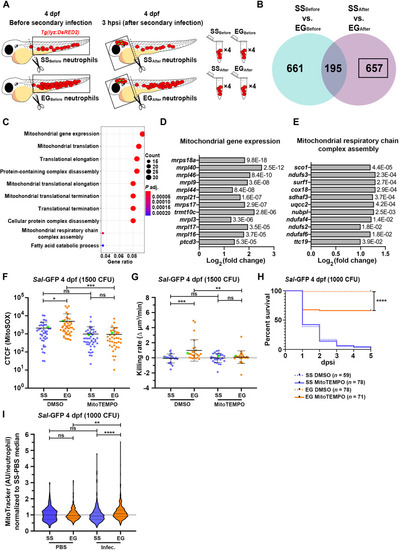
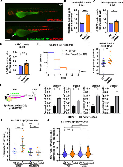
EG neutrophils enhance expression of mitochondria-associated genes following infection, use mtROS for their enhanced bactericidal activity and have greater mitochondrial mass.

(A) Schematic illustrating sample collection for RNA-seq of SS and EG neutrophils before (cells harvested from dissected trunks) and after (cells harvested from dissected heads) infection. (B) Venn diagram of DEGs up-regulated in EG neutrophils that were unique to the “SSBefore versus EGBefore” and “SSAfter versus EGAfter” pairwise comparisons and those common to both comparisons. Black box highlights DEGs of interest. (C) GO:BP analysis of DEGs of interest. (D) Log2 fold change of genes associated with the “mitochondrial gene expression” GO term, with adjusted P values. (E) Log2 fold change of genes associated with the “mitochondrial respiratory chain complex assembly” GO term, with adjusted P values. (F) Quantification of mtROS within Sal-GFP-laden SS and EG neutrophils in the presence of MitoTEMPO or DMSO (control), as detected by MitoSOX fluorescence. Green data points highlight mtROS within neutrophils as shown in fig. S5A. (G) Bacterial killing rates of SS and EG neutrophils in the presence of MitoTEMPO or DMSO (control) following Sal-GFP infection. Green data points highlight killing rates of neutrophils as shown in fig. S5B. (H) Kaplan-Meier graph showing survival of DMSO- and MitoTEMPO-treated SS and EG larvae over 5 dpsi with Sal-GFP at 4 dpf. (I) Flow quantification of mitochondrial mass [as measured with MitoTracker relative fluorescence intensity in arbitrary units (AU)/neutrophil] within individual neutrophils from SS and EG larvae, following Sal-GFP infection at 4 dpf, compared to PBS-injected controls. Error bars, mean ± SD; *P < 0.05, **P < 0.01, ***P < 0.001, ****P < 0.0001; one-way ANOVA with Tukey’s multiple comparisons test (F, G, and I) and Gehan-Breslow-Wilcoxon test (H).

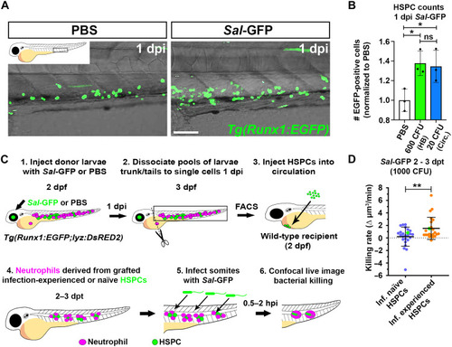
Infection-experienced HSPCs generate neutrophils with elevated bactericidal activity.

(A) Live confocal imaging of HSPCs in the CHT of 3 dpf Tg(Runx1:EGFP) larvae, 1 day following injection with PBS or Sal-GFP. (B) Flow quantification of HSPCs from dissected trunks of Tg(Runx1:EGFP) larvae 1 day following injection with PBS or Sal-GFP into the hindbrain ventricle (HB) or circulation (Circ.), n = 20 larvae per sample, three experimental replicates. (C) Schematic illustrating transplantation strategy to live image neutrophils derived from infection-naïve and infection-experienced HSPCs. (D) Bacterial killing rates of neutrophils derived from infection-naïve and infection-experienced HSPCs. Green data points highlight killing rates of neutrophils as shown in fig. S6H. Error bars, mean ± SD; *P < 0.05, **P < 0.01; one-way ANOVA with Tukey’s multiple comparisons test (B) and unpaired Student’s t test (D). Scale bar, 50 μm.

Larvae with cebpb-overexpressing HSPCs demonstrate enhanced granulopoiesis, survival to infection, and have neutrophils with enhanced bactericidal activity and mitochondrial mass.

(A) Live imaging of Tg(lyz:DsRED2) and Tg(Runx1:cebpb-CG2;lyzDsRED2) larvae at 3 dpf. (B) Flow quantification of neutrophils from 3 dpf Tg(lyz:DsRED2)/WT and Tg(Runx1:cebpb-CG2;lyzDsRED2) larvae (n = 10 larvae per sample, three experimental replicates). (C) Flow quantification of macrophages from the dissected trunks of 3 dpf Tg(mpeg1:EGFP)/WT and Tg(Runx1:cebpb-CG2;mpeg1:EGFP) larvae (n = 10 larvae per sample, three experimental replicates). (D) Flow quantification of HSPCs from the dissected trunks of 3 dpf Tg(Runx1:EGFP)/WT and Tg(Runx1:cebpb-CG2;Runx1:EGFP) larvae, respectively (n = 25 to 50 larvae per sample, three experimental replicates). (E) Kaplan-Meier graph showing survival of Tg(lyz:DsRED2)/WT and Tg(Runx1:cebpb-CG2;lyzDsRED2) larvae over 5 dpsi with Sal-GFP at 3 dpf. (F) Bacterial killing rates of neutrophils within 3 dpf Tg(lyz:DsRED2)/WT and Tg(Runx1:cebpb-CG2;lyzDsRED2) larvae. Green data points highlight killing rates of neutrophils as shown in fig. S7C. (G) Schematic illustrating strategy to FACS-isolate neutrophils from infected Tg(Runx1:cebpb-CG2;lyzDsRED2) larvae. (H) Expression of ndufs3, uqcc2, ndufs2, and ndufaf4 within neutrophils FACS-isolated from infected Tg(lyzDsRED2)/WT and Tg(Runx1:cebpb-CG2;lyzDsRED2) larvae [as shown in (G)], as detected by qPCR (in biological triplicate). (I) Bacterial killing rates of neutrophils within 3 dpf Tg(lyz:DsRED2)/WT and Tg(Runx1:cebpb-CG2;lyzDsRED2) larvae following MitoTEMPO and DMSO (control) treatment. Green data points highlight killing rates of neutrophils as shown in Fig. S7D. (J) Flow quantification of mitochondrial mass (as measured with MitoTracker relative fluorescence intensity in arbitrary units/neutrophil) within individual neutrophils from Tg(lyz:DsRED2)/WT and Tg(Runx1:cebpb-CG2;lyzDsRED2) larvae following Sal-GFP infection at 3 dpf, compared to PBS-injected controls. Error bars, mean ± SD; *P < 0.05, **P < 0.01, ***P < 0.001, ****P < 0.0001; unpaired Student’s t test (B, C, D, F, and H), Gehan-Breslow-Wilcoxon test (E), and one-way ANOVA with Tukey’s multiple comparisons test (I and J). Scale bar, 250 μm.

Acknowledgments
This image is the copyrighted work of the attributed author or publisher, and ZFIN has permission only to display this image to its users. Additional permissions should be obtained from the applicable author or publisher of the image. Full text @ Sci Adv