FIGURE SUMMARY
Title

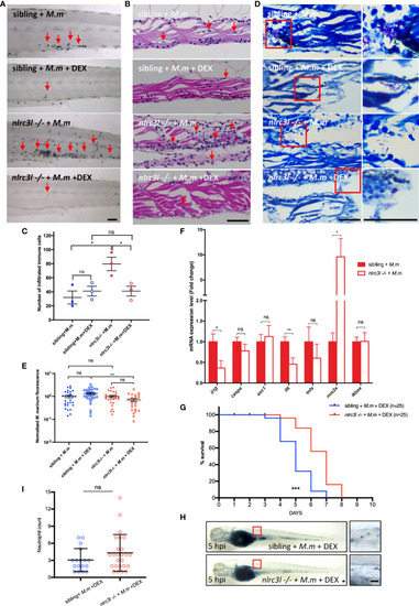
Negative Regulator Nlrc3-like Maintain the Balanced Innate Immune Response During Mycobacterial Infection in Zebrafish

Authors
Niu, L., Luo, G., Liang, R., Qiu, C., Yang, J., Xie, L., Zhang, K., Tian, Y., Wang, D., Song, S., Takiff, H.E., Wong, K.W., Fan, X., Gao, Q., Yan, B.
Source
Full text @ Front Immunol

nlrc3-like deficiency promotes host control of the mycobacterial proliferation during early infection. (A) Zebrafish embryo infections and bacterial fluorescence intensity. The red arrow indicates the duct of Cuvier, the site where bacteria were injected. (B) Representative images for M. marinum:Wasabi infected embryos at 3 dpi. (C) Statistics of relative green fluorescence intensity at the indicated time points taken from two independent biological replicates. (D) Process for calculating granuloma-like structure in zebrafish embryos: i original image; ii. the image after fluorescence-pixelation processing – the area of ​​the red pixels represents the relative size of each granuloma; iii. Calculation of the area of ​​the red pixels. (E) Graph showing the number of large, small and all granuloma-like structures in nlrc3-like -/- and control embryos. (B, D) Scale bar = 200 μm *p ≤ 0.05; **p ≤ 0.01; ns, not statistically significant (p>0.05)..

nlrc3-like deficiency upregulates the expression of inflammatory genes during mycobacterial infection. (A) Quantitative RT-PCR showing expression of nlrc3-like in 3 dpf zebrafish embryos at 0, 1, 3, 5 and 7 days post infection with M. marinum:Wasabi.(B) Quantitative RT-PCR showing expression of multiple inflammatory genes at 1 dpi. The values shown represent an average of three independent biological experiments, 25 embryos per group in each experiment. (C)il-1β WISH shows significantly increased il-1β expression in nlrc3-like deficient mutants compared with their siblings at 1 dpi. Scale bar = 200 μm. (D) Co-staining of il-1β WISH (red) and anti-GFP antibody (green). White arrows point to co-staining signals of mfap4-GFP and il-1β, and white arrow heads point to the il-1β only signals. Scale bar = 50 μm **p ≤ 0.01; ***p ≤ 0.001; ns, not statistically significant (p>0.05).

The impact of nlrc3-like on host defense against mycobacterial infection was mainly mediated by innate immune cells. (A) Schematic view of the construction of nlrc3-like overexpressing embryos and the M. marinum infection assay. pTol2 plasmids were injected at the single-cell stage and subsequently the grown embryos were infected via the duct of Cuvier at 3 dpf. Injections of pTol2-β-actin-nlrc3-like (β-actin-nlrc3l) and pTol2-coronin1a-nlrc3-like (coro1a-nlrc3l) generated embryos overexpressing nlrc3-like in all cells or only in myeloid cells, respectively. Black arrows indicate injection sites. (B) Graph of the relative green fluorescence intensity of the β-actin-nlrc3l group and the β-actin-6xmyc group at the indicated time points. (C) Graph of the relative green fluorescence intensity of the coro1a-nlrc3l and coro1a-6xmyc embryos at the indicated time points. (D) Graph showing the number of granuloma-like structures in the coro1a-nlrc3l and the coro1a-6xmyc embryos. (E) Survival curve of coro1a-nlrc3l and coro1a-6xmyc embryos. The data shown represents the averages from two independent biological replicates *p ≤ 0.05; **p ≤ 0.01; ***p ≤ 0.001; ns, not statistically significant (p>0.05).

nlrc3-like deficiency promotes the activation of macrophage inflammasome. (A) Schematic view of FACS of mfap4-GFP+ macrophages with phagocytosed Tdtomato+M. marinum and subsequent detection of the expression of inflammation-related genes by RT-PCR. (B) A representative image of zebrafish embryos infected with M. marinum:Tdtomato at 10 hpi. i uninfected Tg (mfap4-GFP) embryo; ii. Tg (mfap4-GFP) embryo infected with M. marinum:Tdtomato; iii. Enlarged view of caudal hematopoietic tissue (CHT) region in ii. white arrows indicate macrophages with phagocytosed bacteria; white triangles indicate macrophages without phagocytosed bacteria; white asterisks, free M. marinum. (C) Gating strategy for FACS. i: Sample: Tg(mfap4: GFP) embryos infected with M. marinum:Tdtomato; ii. uninfected wild-type zebrafish; iii. Tdtomato single color control, wild-type zebrafish infected with M. marinum:Tdtomato; iv. GFP single color control, uninfected Tg(mfap4: GFP). P1, free M. marinum:Tdtomato; P2, mfap4-GFP+ macrophages; P3, mfap4-GFP+ macrophages with phagocytosed M. marinum:Tdtomato. (D) Expression of pro-inflammatory genes in macrophages at 10 hpi. The values represent the average of three independent biological repeats, 25 embryos per group in each repeat. (E) The expression of pro-inflammatory genes in embryos with overexpressing nlrc3-like in myeloid cells. (F) Representative images of M. marinum:wasabi proliferation in nlrc3-like -/-, asc -/- nlrc3-like -/-, and their nonmutant siblings. (G) Graph of green fluorescence intensity in nlrc3-like -/-, asc -/- nlrc3-like -/-, and their nonmutant siblings at the indicated time points. The values shown are the combined results of two independent experimental replicates. (B, F) scale bar = 200 μm *p ≤ 0.05; **p ≤ 0.01; ***p ≤ 0.001; ns, not statistically significant (p>0.05).

nlrc3-like deficiency promotes neutrophil recruitment. (A) Survival curves of nlrc3-like -/- and non-mutant embryos after M. marinum infection, based on results from two independent biological replicates. (B) Numbers of neutrophils recruited to infection sites at the indicated time points. (C) Live imaging of neutrophils after M. marinum infection. 3 dpf Tg (lyz-GFP) zebrafish embryos were infected with M. marinum:Tdtomato. Time interval, 1 min 30 s. The data is representative of three independent biological replicates. (D) SB staining showing neutrophil recruitment to infection sites. The left panel shows the whole embryo while the right panel shows magnification of the area in the red box used for counting neutrophils. (E) Graph of neutrophils counted in (D, F) Graph of the neutrophils counted after SB staining of β-actin-nlrc3l (whole body expression of nlrc3-like) and β-actin-6Xmyc embryos. (G) Graph of neutrophils counted after SB staining in coro1a-nlrc3l (myeloid expression of nlrc3-like) and coro1a-6Xmyc embryos (C, D). Scale bar = 50 μm *p ≤ 0.05; **p ≤ 0.01; ***p ≤ 0.001.

nlrc3-like deficiency leads to excessive neutrophils infiltration and tissue damage at later stages of infection. (A) SB staining of the tail region in different groups of zebrafish embryos. (B) H&E staining of paraffin sections of the caudal vein plexus. (C) Quantification of the number of infiltrated immune cells in (B, D) Acid fast staining of paraffin sections in the caudal vein plexus. (E) Statistics of normalized bacterial fluorescence intensity at late stage of infection. (F) Quantitative RT-PCR assessed the expression of multiple cytokines at a late stage of infection (6 dpi). The values are averages from three independent biological repeats, 25 embryos per group in each repeat. (G) Survival curves of infected zebrafish with DEX treatment. The data is from one of two similar independent biological replicates (H). SB staining showing neutrophil recruitment to infection sites at 5 hpi. The left panel shows the whole embryo while the right panel shows an enlargement of the region in the red box that was used for counting neutrophils. (I) Graph showing the number of neutrophils, counted as in (H). The data shown was combined from three independent biological repeats. (A, B, D, H) Scale bar = 50 μm *p ≤ 0.05; **p ≤ 0.01; ***p ≤ 0.001; ns, not statistically significant (p>0.05).

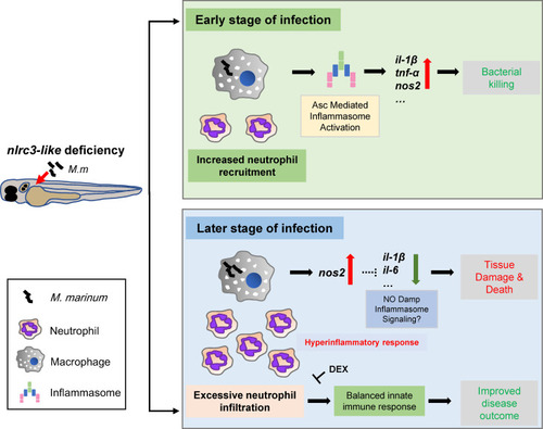
Model for the impact of negative regulator NLR nlrc3-like on innate immune responses during mycobacterial infection. During the early stage of infection, the defect of nlrc3-like can boost the expression of mycobacterial elicited pro-inflammatory genes via Asc mediated inflammasome activation in infected macrophages, which results in a bacterial killing effect accompanied by an increased neutrophil recruitment. However, during the late stage of infection the host shifts into a hyperinflammatory status with uncontrolled excessive neutrophil infiltration and increased tissue damage and death. The administration of DEX can block the excessive inflammatory response and improve disease outcome.

Acknowledgments
This image is the copyrighted work of the attributed author or publisher, and ZFIN has permission only to display this image to its users. Additional permissions should be obtained from the applicable author or publisher of the image. Full text @ Front Immunol