FIGURE SUMMARY
Title

In vivo Evaluation of Non-viral NICD Plasmid-Loaded PLGA Nanoparticles in Developing Zebrafish to Improve Cardiac Functions

Authors
Messerschmidt, V.L., Chintapula, U., Bonetesta, F., Laboy-Segarra, S., Naderi, A., Nguyen, K.T., Cao, H., Mager, E., Lee, J.
Source
Full text @ Front. Physiol.

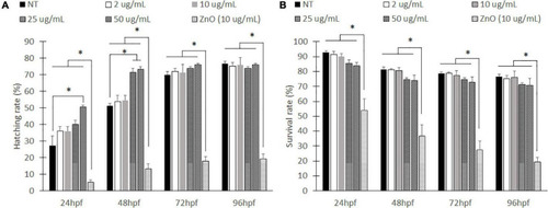
Hatching rates and survival rate for embryonic zebrafish toxicity study. Hatched to unhatched ratio (A) and survival rate (B) of non-treated fish to various concentrations of empty PLGA nanoparticle suspension. *indicates a significant difference (p < 0.01). n = 50 fish per group.

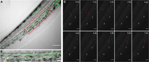
Coumarin-6 Loaded Nanoparticles after Injection. (A) Coumarin-6 nanoparticles traveling in the caudal artery of the zebrafish tail. Red box is blown up and shown as (B). White arrows show nanoparticles rolling along vessel walls. Yellow arrows indicate clusters of endocytosed nanoparticles. (C) Representative stills showing nanoparticles traveling from the caudal artery (0.2–1.2 s), into the capillaries of the tail tissue (1.6–4.8 s) and entering the posterior caudal vein (6.6–7.4 s). All scale bars represent 100 μm.

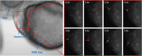
Coumarin-6 Nanoparticles Rolling on Endocardium. Representative stills showing nanoparticles flowing through the cardiac chambers. Arrows indicate injected nanoparticle. All scale bars represent 100 μm.

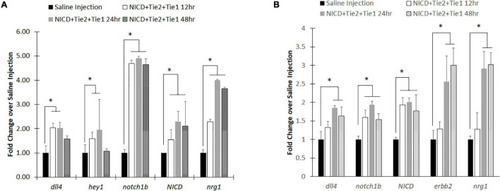
RT-PCR analysis after injecting intravenous NICD loaded nanoparticles. (A) Notch signaling components were all upregulated after injection of NICD loaded nanoparticles until 24 h post injection. However, Notch ligand (dll4) and receptor (notch1b) were returned to control group (saline injection) level while downstream components (nrg1 and hey1) remain high with NICD expression level. (B) Isolated zebrafish heart after injection showed higher expression levels of Notch components to increase cardiac function. Interestingly, endocardial nrg1-myocardial erbb2 signaling components consistently increased up to 48 h post injection. * indicates a significant difference (p < 0.01). n = 5.

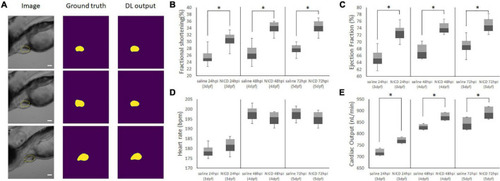
Representative Images of Model Output for analyzing cardiac functions. (A) Representative images of the image input (column 1), hand segmented cardiac volume (column 2), and the model’s predicted cardiac volume after training the ZACAF network (column 3). (B,C) FS and EF comparisons demonstrated the zebrafish heart contracts stronger after injecting NICD incorporated PLGA nanoparticles. (D) Injection of NICD loaded PLGA nanoparticles didn’t affect heart rate. (E) Cardiac output of both control group and NICD loaded nanoparticles gradually increased as zebrafish heart matures. However, nanoparticles injected zebrafish has higher number of cardiac output. * indicates a significant difference (p < 0.01). n = 250 zebrafish images per group.

Acknowledgments
This image is the copyrighted work of the attributed author or publisher, and ZFIN has permission only to display this image to its users. Additional permissions should be obtained from the applicable author or publisher of the image. Full text @ Front. Physiol.