
- Title
-
Zebrafish duox mutations provide a model for human congenital hypothyroidism
- Authors
- Chopra, K., Ishibashi, S., Amaya, E.
- Source
- Full text @ Biol. Open

duox mutants exhibit growth retardation. Mutants for both alleles as well as compound heterozygotes are shorter than their WT and heterozygous siblings at 3 months (A–G) but catch up by 6 months (H). sa13017−/− animals are trailing behind even at 6 months (H). Asterisks in G denote statistically significant differences (Bonferroni's multiple comparisons test, ****P<0.0001) duox mutants also have a delay in the inflation of the anterior lobe of the swim bladder (I,J) (white arrowheads indicate lobes). Adults at 3 months old also lack barbels (L–P), which are observed in heterozygous siblings (white arrowheads; K,N). Barbels emerge in some older animals (6 months and older) (white arrowhead, M). External goitres are often visible in young adults (black arrowheads; L,O). Scale bars: 1 mm. PHENOTYPE:
|

Adult duox mutant zebrafish display an array of visible phenotypes. (A–C) 5× magnification of flank region showing the distribution of melanophores in WT, sa9892+/− and sa989−/− siblings. The apparent abundance of melanophores was statistically significant in duoxmutants (D). Asterisks denote statistically significant differences (Bonferroni's multiple comparisons test, ****P<0.0001). duox mutants also showed irregularities in stripe pattern in contrast to heterozygous siblings, shown here in a 2× magnification of the flank in sa9892siblings (E,F). Craniofacial anomalies were evident among mutants, with frontal height significantly shorter among mutants (G–I) (Bonferroni's multiple comparisons test, *P<0.5, **P<0.01). Erythema in the thoracic region was prominent among mutants. This was especially noticeable in nacre backgrounds (L,N,O). duox mutants also suffered from perpetual fin damage, which manifest as ragged margins and tears (S–U). Scale bars: 1 mm. |
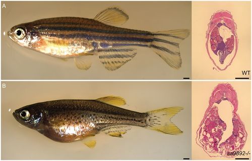
duox mutant females are unable to ovulate and become egg bound. H&E staining of abdominal sections reveals oocytes (A,B). Scale bars: 1 mm. PHENOTYPE:
|
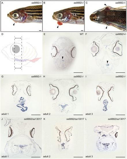
Homozygous duox mutations lead to goitre. Adult mutant animals exhibit an array of variably sized external goitres (arrowheads; A,B), as well as lateral flaring of opercula (arrowheads; C). When sectioned along the length of the follicular region (dotted area, D) and subjected to ISH for thyroglobulin, mutants reveal extensive spread of and ectopic thyroid follicular tissue (G–L), in contrast to the localised, discreet distribution in WT and heterozygous siblings (arrowheads; E,F). Scale bars: 1 mm. |
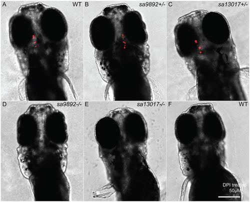
Hypothyroidism is evident among duox mutants. At 5 dpf, homozygous mutant larvae lack staining for bound T4 in the thyroid follicles, based on wholemount fluorescent immunohistochemistry (D,E). This is in sharp contrast to the robust staining observed in WT and heterozygous siblings (A–C). The NADPH oxidase inhibitor DPI successfully phenocopies duoxmutations in WT larvae, resulting in an absence of T4 detection (F). Scale bar: 50 μm. |

T4 treatment alleviates phenotypic anomalies in duox mutants. T4-treated mutants show an improvement in fin health, compared to untreated mutants (A,B). Pigment changes are evident among T4-treated mutants. C–F show a 5× magnification of the distribution of melanophores on the flank region of sa9892+/− and sa989−/− siblings, with a significant reduction in melanophore number (G). Asterisks denote statistically significant differences (Bonferroni's multiple comparisons test, ****P<0.0001). Goitres resolve following T4 administration, but small ectopic thyroids are still evident (black arrowheads) (I,J). Scale bars: 1 mm. |

The goitrogen methimazole (MMI) phenocopies duox mutations. A and B show a 5× magnification of the distribution of melanophores on the flank region among MMI-treated and untreated WT fish. Treated animals have at least two distinct populations of melanophores, based on size (A,B). Pigment change pertaining to melanophore numbers is significant following MMI treatment (C). (Bonferroni's multiple comparisons **P<0.01). MMI leads to loss of bound T4in WT larvae (D,E) and induces external goitre (arrowhead; F). ISH for thyroglobulin reveals widespread follicular tissue, not limited to the mid-ventral region (H), similar to duox mutants (I). Scale bars: 1 mm. |