
|
Fig. 8
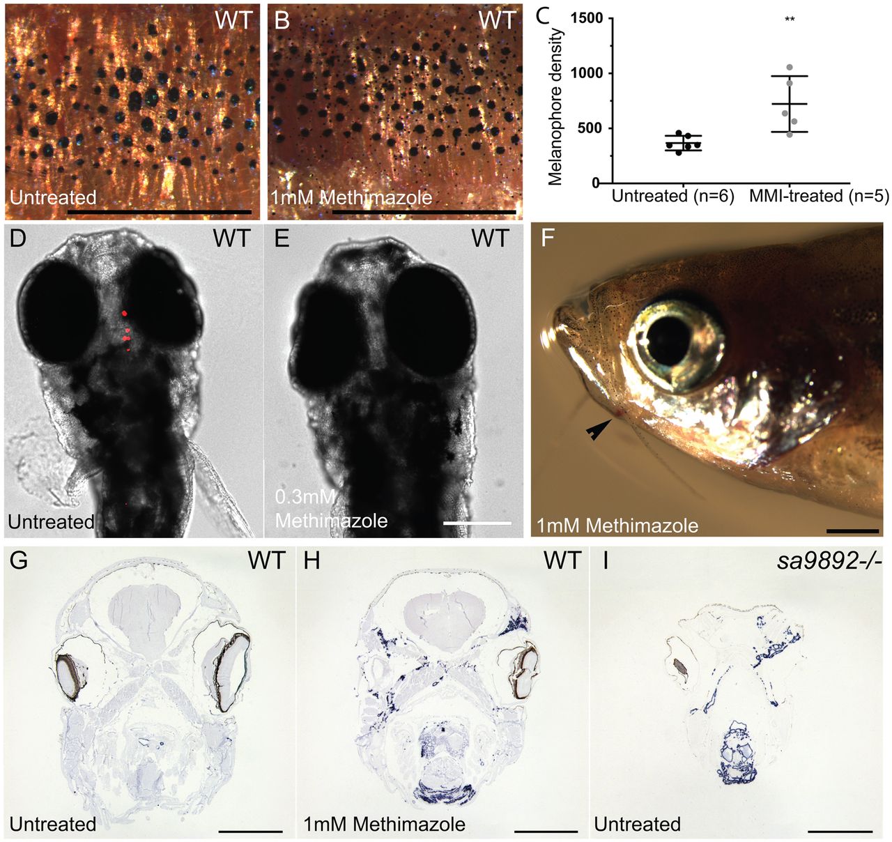
The goitrogen methimazole (MMI) phenocopies duox mutations. A and B show a 5× magnification of the distribution of melanophores on the flank region among MMI-treated and untreated WT fish. Treated animals have at least two distinct populations of melanophores, based on size (A,B). Pigment change pertaining to melanophore numbers is significant following MMI treatment (C). (Bonferroni's multiple comparisons **P<0.01). MMI leads to loss of bound T4in WT larvae (D,E) and induces external goitre (arrowhead; F). ISH for thyroglobulin reveals widespread follicular tissue, not limited to the mid-ventral region (H), similar to duox mutants (I). Scale bars: 1 mm.