
- Title
-
Generation of motor neurons requires spatiotemporal coordination between retinoic acid and Mib-mediated Notch signaling
- Authors
- Kong, H.J., Ryu, J.H., Kim, J., Kim, J.W., Seong, B., Whang, I., Park, J.Y., Yeo, S.Y.
- Source
- Full text @ Animal Cells Syst (Seoul)
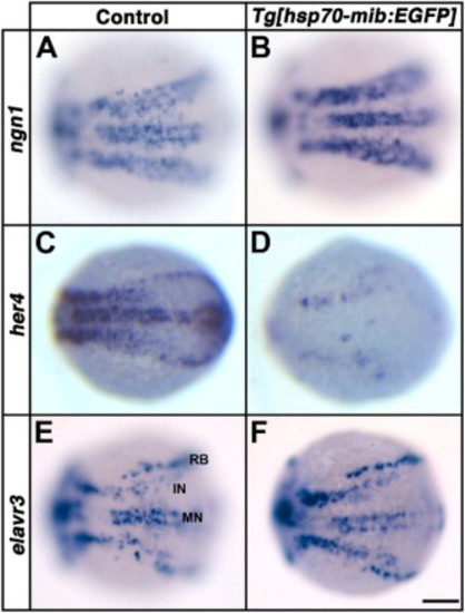
Overexpression of Mib:EGFP causes a reduction in Notch signaling. Dorsal views. Anterior to the left. The expression of ngn1 (A, B), her4 (C, D) and elarvr3 (E, F) in control (A, C and E) and heterozygous Tg[hsp70-mib:EGFP] embryos at 11 hpf (B, D and F) following heat-shock at 6 hpf. RB, Rohon-Beard neuron; IN, interneuron; MN, motor neuron. Scale bar: 100 µm. |
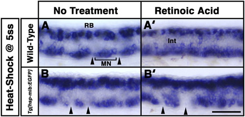
Overexpression of Mib:EGFP attenuates the effect of exogenous retinoic acid. Lateral views. Anterior to the left. At 26 hpf, expression of islet1 between the 8th and 11th somites of embryos following no treatment (A and B) and retinoic acid (RA) treatment (A’ and B’). Wild-type (A and A’) and Tg[hsp70-mib:EGFP] (B and B’) embryos were heat-shocked at the 5-somite stage (ss). Motor neuron (MN) clusters (brackets) located in a somite flanking a non-MN domains (arrowhead) in wild-type and Tg[hsp70-mib:EGFP] embryos. Scale bar: 100 µm. |
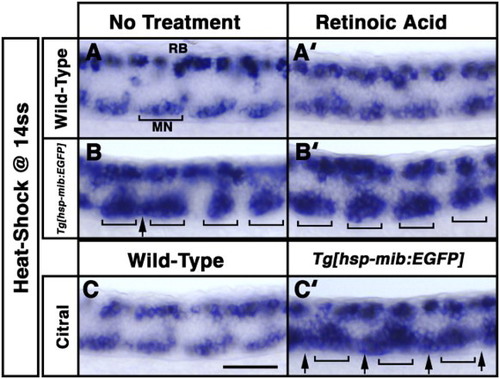
Retinoid pathway attenuates the effect of overexpression of Mib:EGFP. Lateral views. Anterior to the left. At 26 hpf, expression of islet1 between the 8th and 11th somites of embryos following no treatment (A–C), retinoic acid (RA) treatment (A’ and B’) or citral treatment (C and C’). Wild-type (A, A’ and C) and Tg[hsp70-mib:EGFP] (B, B’ and C’) embryos were heat-shocked at the 14-somite stage (ss). Motor neuron (MN) clusters (brackets) located in the somites flanking non-MN domain in wild-type embryos. Ectopic motor neurons (arrows) filled the gaps between motor neuron clusters (brackets) in Tg[hsp70-mib:EGFP] embryos following heat-shock at the 14ss (B) and exposure to citral at the 14ss (C’). Scale bar: 100 µm. |
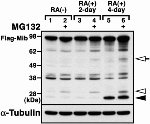
Proteolysis of Mib in the presence of retinoic acid. Flag-tagged Mib (Flag-Mib) was transfected into P19 cells (lanes 1 and 2), P19 cells treated with retinoic acid (RA) for 2 days (lanes 3 and 4) or P19 cells treated with RA for 4 days (lanes 5 and 6) in the absence or presence of MG132. Western blot analysis using the anti-Flag antibody revealed the proteolytic fragments of Mib. Arrowhead and open arrowhead indicates the retinoid pathway-dependent 27 kDa fragments and proteasome-dependent 30 kDa fragments, respectively. Open arrows indicate the proteolytic fragments, depending on both the retinoid pathway- and proteasome-dependent 55 kDa fragments. |