
|
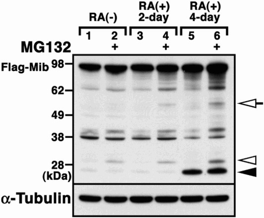
Fig. 4 Proteolysis of Mib in the presence of retinoic acid. Flag-tagged Mib (Flag-Mib) was transfected into P19 cells (lanes 1 and 2), P19 cells treated with retinoic acid (RA) for 2 days (lanes 3 and 4) or P19 cells treated with RA for 4 days (lanes 5 and 6) in the absence or presence of MG132. Western blot analysis using the anti-Flag antibody revealed the proteolytic fragments of Mib. Arrowhead and open arrowhead indicates the retinoid pathway-dependent 27 kDa fragments and proteasome-dependent 30 kDa fragments, respectively. Open arrows indicate the proteolytic fragments, depending on both the retinoid pathway- and proteasome-dependent 55 kDa fragments.