
- Title
-
The Vital Dye CDr10b Labels the Zebrafish Mid-Intestine and Lumen
- Authors
- Sander, V., Patke, S., Lee, J.Y., Chang, Y.T., Davidson, A.J.
- Source
- Full text @ Molecules
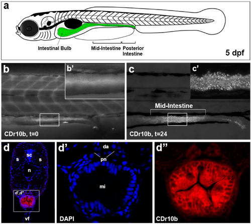
CDr10b labels the mid-intestine. (a) Schematic of a 5 dpf larva with the gut in green; (b,b’) After treatment (100 nM, 45 min), CDr10b is found in the blood and gut lumen (t = 0); (c) 24 h post treatment (t = 24), cells of the mid-intestine are stained by CDr10b; (c’) Localization of CDr10b in a punctate-pattern in enterocytes; (d–d’’) Transverse section through the mid-intestinal segment showing CDr10b staining (red in d) and nuclear DAPI staining (blue in d). da, dorsal aorta; dpf, days post fertilization; mi, mid-intestine; n, notochord; pn, pronephros; t, time post treatment; s, somites; sc, spinal cord; vf, ventral fin. |
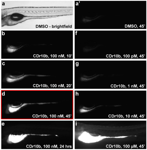
Dosage and duration of CDr10b treatment. Larvae were treated at 5 dpf with 0.01% DMSO or CDr10b at the doses and duration indicated in the panels, and photographed 24 h post treatment (6 dpf). (a) Brightfield image of a 5 dpf control (DMSO-treated) larva. (a') Same specimen photographed using a red filter set. Only background fluorescence is detectable; (b–e) Increasing incubation times with constant dosage (100 nM CDr10b); (f–i) Increasing dosage of CDr10b with constant incubation time (45 min). All images were captured using the same exposure settings. Red box: Optimal conditions for CDr10b staining. For survival rates see Table S1. ', minutes; hrs, hours; t, time post treatment. |
|
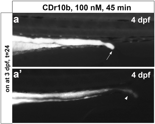
Stages of CDr10b staining. (a,a') CDr10b taken up at 3 dpf stays in the intestinal lumen and can be used to visualize the timing of cloaca opening. Arrow in a, closed cloaca. (b) CDr10b treatment at 4 dpf results in luminal staining with excretion through the cloaca (arrowheads in a' and b); (c) CDr10b treatment at 5 dpf leads to uptake of the dye specifically by mid-intestinal epithelial cells; (d) The CDr10b signal in the mid-intestine is stable 4 days post treatment (t = 96). dpf, days post fertilization; t, time post treatment. 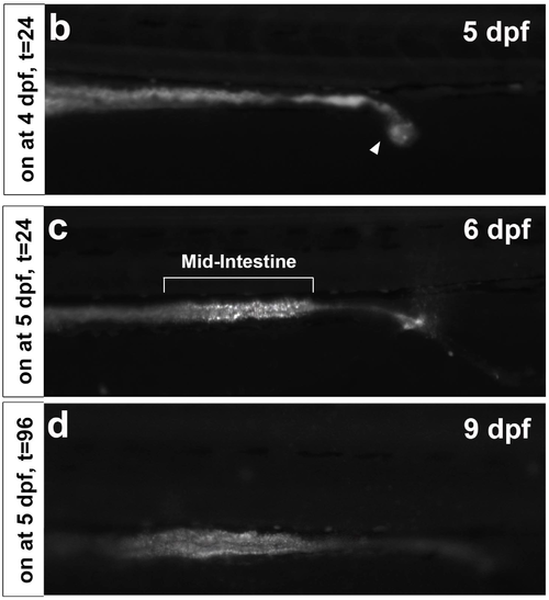
|
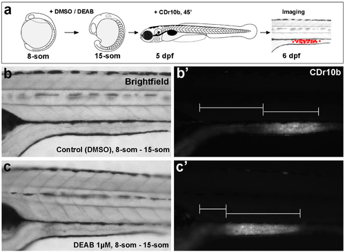
CDr10b labels developmental malformation of the intestine. (a) Outline of the experiment; (b,c) Brightfield images reveal that diethlyaminobenzaldehyde (DEAB) treatment during early development does not affect the morphology of the larvae; (b',c') Detection of the CDr10b signal reveals that the mid-intestinal segment is shifted towards anterior upon retinoic acid (RA) inhibition. |
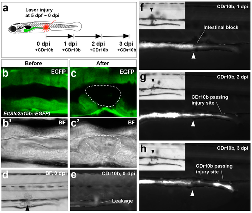
CDr10b as a readout for intestinal injury and regeneration. (a) Outline of the experiment; (b–c') Confocal live imaging of mid-intestinal epithelium of a Et(Slc2a15b::EGFP) larva before and after laser cell ablation. Detection of the green fluorescence protein (GFP) signal (b,c) and the corresponding brightfield view (b',c'). Intact epithelium (b,b'). After laser exposure, the cells have lost GFP expression and appear necrotic (white outline in c and c'); (d,e) Immediately after injury (0 dpi), the intestinal tube is partially blocked (brightfield in d) and CDr10b leaks into the surrounding tissue (CDr10b signal in e); (f–h) The same fish photographed at 1, 2 and 3 dpi after CDr10b treatment shows the flow of CDr10b blocked at 1 dpi, and regeneration with successful excretion of CDr10b by 3 dpi. Arrowheads mark the site of injury. BF, brightfield; dpi, day(s) post injury. |