
|
Fig. 1
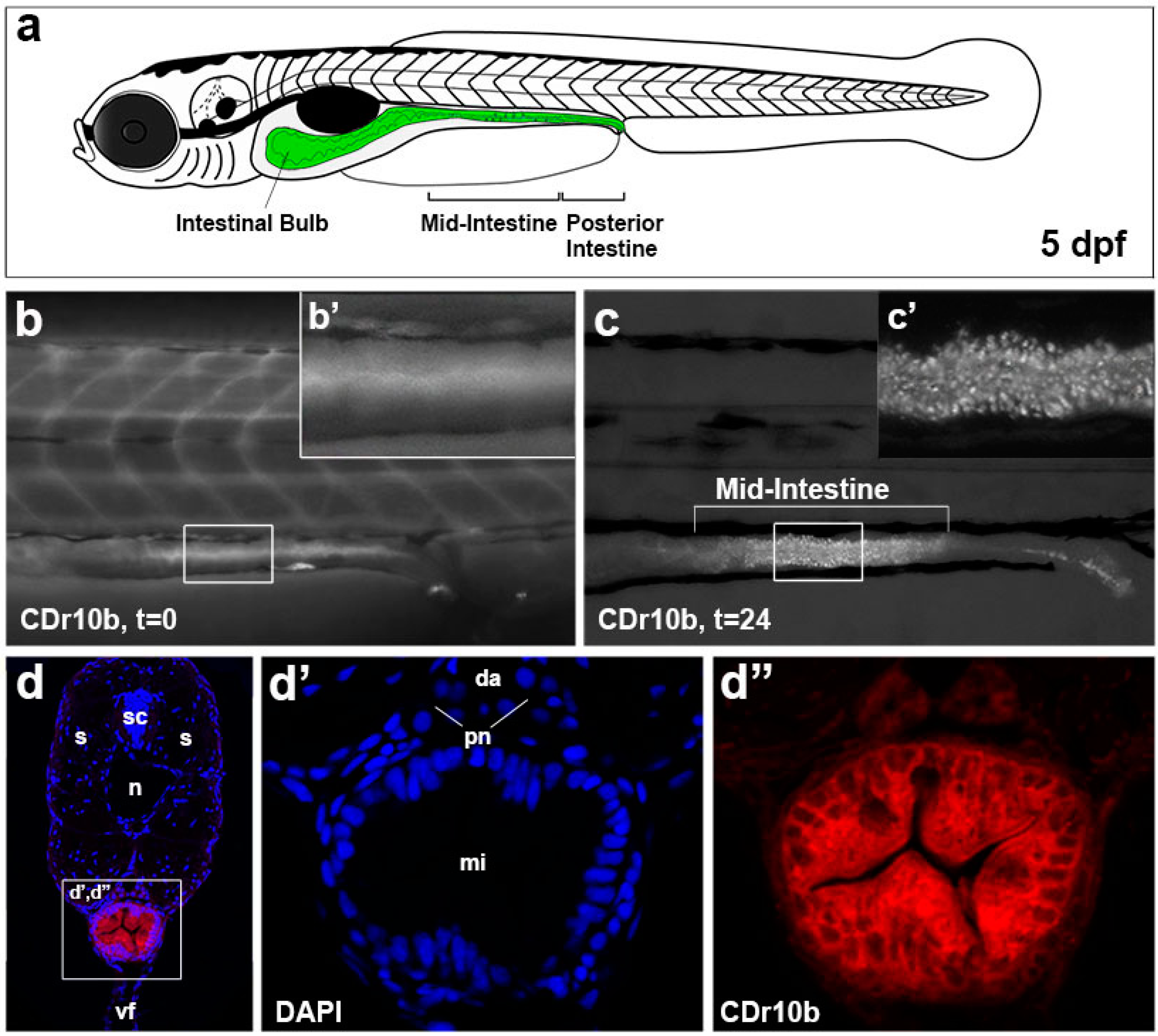
CDr10b labels the mid-intestine. (a) Schematic of a 5 dpf larva with the gut in green; (b,b’) After treatment (100 nM, 45 min), CDr10b is found in the blood and gut lumen (t = 0); (c) 24 h post treatment (t = 24), cells of the mid-intestine are stained by CDr10b; (c’) Localization of CDr10b in a punctate-pattern in enterocytes; (d–d’’) Transverse section through the mid-intestinal segment showing CDr10b staining (red in d) and nuclear DAPI staining (blue in d). da, dorsal aorta; dpf, days post fertilization; mi, mid-intestine; n, notochord; pn, pronephros; t, time post treatment; s, somites; sc, spinal cord; vf, ventral fin.