
|
Fig. 5
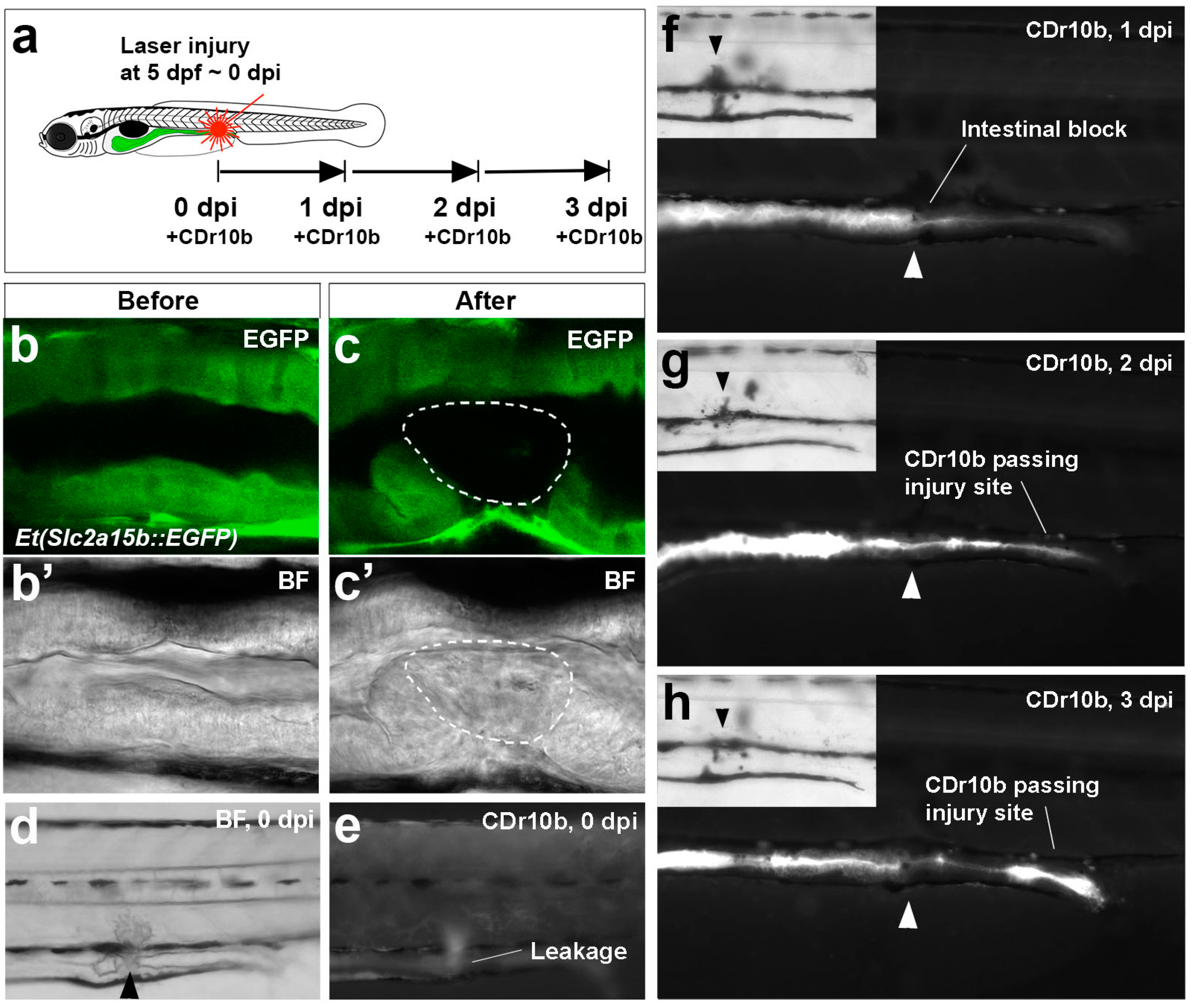
CDr10b as a readout for intestinal injury and regeneration. (a) Outline of the experiment; (b–c') Confocal live imaging of mid-intestinal epithelium of a Et(Slc2a15b::EGFP) larva before and after laser cell ablation. Detection of the green fluorescence protein (GFP) signal (b,c) and the corresponding brightfield view (b',c'). Intact epithelium (b,b'). After laser exposure, the cells have lost GFP expression and appear necrotic (white outline in c and c'); (d,e) Immediately after injury (0 dpi), the intestinal tube is partially blocked (brightfield in d) and CDr10b leaks into the surrounding tissue (CDr10b signal in e); (f–h) The same fish photographed at 1, 2 and 3 dpi after CDr10b treatment shows the flow of CDr10b blocked at 1 dpi, and regeneration with successful excretion of CDr10b by 3 dpi. Arrowheads mark the site of injury. BF, brightfield; dpi, day(s) post injury.