
- Title
-
Distinct expression patterns of syndecans in the embryonic zebrafish brain
- Authors
- Hofmeister, W., Devine, C.A., and Key, B.
- Source
- Full text @ Gene Expr. Patterns
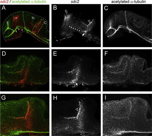
sdc2 expression in zebrafish neuroepithelium. Confocal sections of the zebrafish brain at 28 hpf, showing expression of sdc2 (B, E, H and in red A, D, G) in relation to the axon scaffold (C, F, I and in green A, D, G). Compiled z-sections are shown in A-C while 2.0 µm sections are represented in D-I. Panels-D-F represent higher magnificantions of A-C at the level of the isthmus with D-F being medial optical slices and G-I more lateral sections. In all panels rostral is to the left and dorsal to the top. Strong expression is particularly seen at the rostraldorsal diencephalon adjacent to the dorsal most point of the forebrain ventricle (asterix in A, unfilled arrow in B) and adjacent to the midbrain ventricle (unfilled arrowhead in B and H), expression in this region extends into the isthmus (dashed yellow outline in A, unfilled arrow in B and H). Weak expression between the diencephalon and tegmentum is also indicated (small filled arrows in B). The telencephalon (T), tectum (Tt) and cerebellum (C) all remain absent for sdc2 expression. Scale bar in C and I is 100 µm. EXPRESSION / LABELING:
|
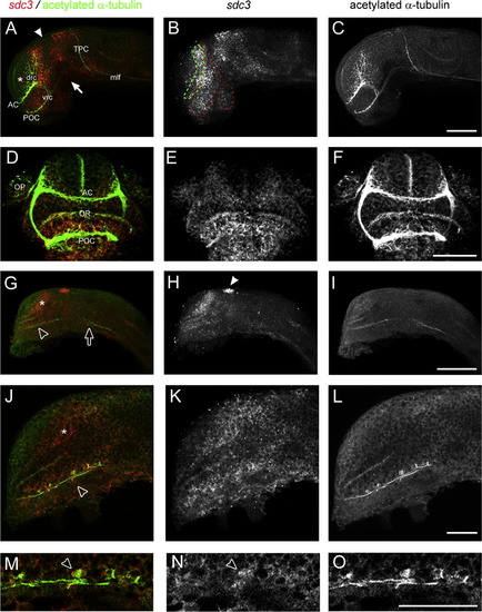
sdc3 expression in the neuroepithelium correlates to regions of neurogenesis and axogenesis. Confocal images of wholemount zebrafish brains at various developmental stages stained for sdc3 expression (B, E, H, K, N and in red A, D, G, J, M) and the pan-axonal marker acetylated tubulin (C, F, I, L, O and in green A, D, G, J, M). Dorsal is to the top and rostral to the left in all panels except (D-F) where rostral is facing. Embryos were analysed at 28 hpf (A-F) and 18 hpf (G-O). In the telencephalon and diencephalon at both 18 and 28 hpf we see a high concentration of sdc3 in the neuroepithelium broadly correlating to regions of the developing axon scaffold. Occasionally an overlap is seen with tubulin stained cells indicating expression in neurons, in this case in neurons of the ventrorostral cluster (M-O) at 18 hpf (unfilled arrowhead in M and N). At 28 hpf staining is strong in the ventral telencephalon (green dashed outline) and ventrodorsal diencephalon (red dotted outline) but absent from the middle of the diencephalon (filled arrowhead in A) rostral dorsal telencephalon (asterix in A) and at the TPOC (filled arrow in A) Abbreviations; anterior commissure (AC), post-optic commissure (POC), optic recess (OR), olfactory placode (OP) dorsorostral cluster (drc), ventrorostral cluster (vrc), tract of posterior commisure (TPC) and medial longitudinal fascicle (mlf). Scale bars in C, F and I are 100 µm, Scale bars in L and O are 25 µm. EXPRESSION / LABELING:
|

The POC is surrounded by sdc4. Compiled confocal sections of wholemount zebrafish brains at 28 hpf stained for sdc4 expression (B, E, H, K, and in red A, D, G, J) and HNK-1 (C, F, I, L and in green A, D, G, J), dorsal to the top and rostral to the left in all panels except (J-L) where rostral is facing. Panels D-F show a high magnification view of the telencephalon and diencephalon and Panels G-I show a high magnification view of the ventrocaudal cluster. Expression is absent from the Telencephalon (T) and Tectum (Tt), being concentrated in the dorsal diencephalon (unfilled arrow in D), isthmus (unfilled arrow in A) and in the rostral diencephalon, in the vicinity of the optic stalk, where it surrounds the post-optic commissure POC (asterix in A, D and J). Abbreviations; anterior commissure (AC), dorsorostral cluster (drc), post-optic commissure (POC), tectum (Tt), tegmentum (Tm), telencephalon (T), ventral diencephalon (vD), ventral caudal cluster (vcc), ventrorostral cluster (vrc). Scalebar in C is 100 µm, scale bar in F, I and L is 25 µm. EXPRESSION / LABELING:
|
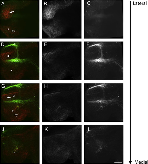
sdc4 expression surrounds the POC at 28 hpf. 2.0 µm optical saggital section taken through wholemounts of the zebrafish forebrain at 28 hpf from most lateral (A-C) to most medial (J-L). sdc4 expression (B, E, H, K and in red A, D, G, J) is shown in relation to the anterior and post-optic commissures and the neuronal clusters of the drc and vrc that contribute to these commissures respectively. Single sections taken laterally reveal the presence of sdc4 positive neuroepithelium lateral to the post-optic commissure (filled arrow in A). More medial sections reveal expression between the optic recess and POC (filled arrow in D-J) and also ventral to it in the presumptive hypothalamus (asterix, D-J). Abbreviations; dorsorostral cluster, optic recess (or), hypothalamus (hy), ventrorostral cluster (vrc). Scalebar in L is 25 µm. EXPRESSION / LABELING:
|
Reprinted from Gene expression patterns : GEP, 13(3-4), Hofmeister, W., Devine, C.A., and Key, B., Distinct expression patterns of syndecans in the embryonic zebrafish brain, 126-32, Copyright (2013) with permission from Elsevier. Full text @ Gene Expr. Patterns