
- Title
-
Tissue and cell-specific transcriptional activity of the human cytomegalovirus immediate early gene promoter (UL123) in zebrafish
- Authors
- Mella-Alvarado, V., Gautier, A., Le Gac, F., and Lareyre, J.J.
- Source
- Full text @ Gene Expr. Patterns
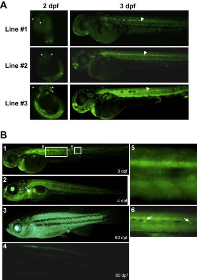
External fluorescence observed in hCMV-IE:eGFP transgenic zebrafish. (A) Transgenic zebrafish were observed with a macroscope equipped with a GFP filter. A similar tissue distribution of transgene expression was observed in three independent transgenic zebrafish lines although different expression levels were observed. Note that transgenic zebrafish lines #1, #2 and #3 showed low, medium and high transgene expression levels, respectively. Transgene was mainly expressed in the neurons of the olfactory placodes (asterisks), the neuromasts, and in the central canal of the spinal cord (arrowheads). (B) Transgene expression pattern during ontogeny: (1) 3 day-old hatched embryo; (2) 4 day-old larvae, the white arrow indicates the liver; (3) 60 day-old transgenic juvenile; (4) 60 day-old wild type (WT) juvenile; (5) inset of 1 showing a high magnification of the spinal cord; (6) inset of 1 showing a high magnification of the lateral neuromasts (white arrows). EXPRESSION / LABELING:
|
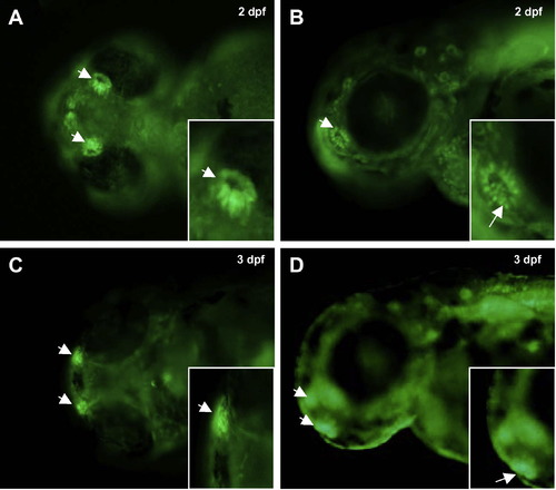
Transgene expression in a subset of cells of the olfactive placodes. Ventral (A and C) and lateral (B and D) views of 2 and 3 dpf transgenic zebrafish larvaes. Note the migration of two symmetric patches of highly fluorescent cells in the olfactory placodes. EXPRESSION / LABELING:
|
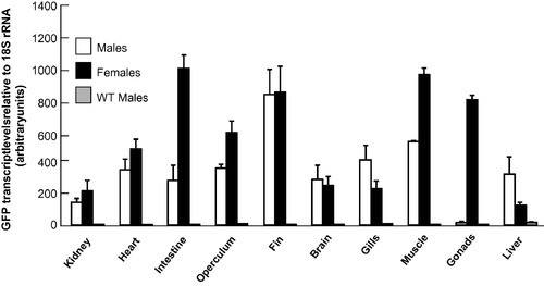
Tissue distribution of the gfp transcripts in adult zebrafish. Transgene expression was assessed by quantitative real time PCR on a panel of tissues collected from male and female adult transgenic zebrafish. Wild type (WT) male were included as a negative reference. gfp mRNA abundance was normalized to 18S rRNA. Transgene expression was detected in all tissues examined with the exception of the testis. |
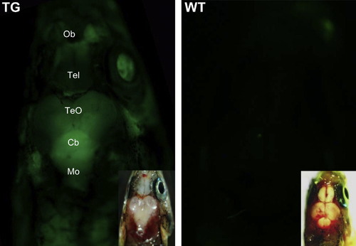
Fluorescence in the brain of hCMV-IE:eGFP transgenic zebrafish. The Highest levels of fluorescence were observed in the regions of the cerebellum of transgenic (TG) but not wild type (WT) zebrafish. The inset shows the brightfield image of the fish. Ob; Olfactory bulb; Tel: Telencephalon, TeO: Optic Tectum; Cb: Cerebellum; Mo: Medulla oblongata. EXPRESSION / LABELING:
|
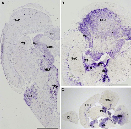
Transgene expression is regionalized in the brain of hCMV-IE:eGFP transgenic lines. The regionalized expression of the gfp transcripts was studied in the brain of adult zebrafish by whole-mount RNA in situ hybridization with a digoxigenin-labeled antisense riboprobe. (A) Cross section of the brain at the level of the midbrain and hindbrain boundary (MHB). (B) Horizontal section of the cerebellum and surrounding area. (C) Sagittal section of the brain. Scale bars represent 500 μm. CCe: Corpus cerebelli; DI: lateral zone of dorsal telencephalic area; DIL: diffuse nucleus of the inferior lobe; DTN: dorsal tegmental nucleus; GC: griseum centralis; MLF: medial longitudinal fascicle; Mo: medulla oblongata; TeO: tectum opticum; TL: torus longitudinalis; TLa: torus Lateralis; TS: torus semicircularis; TPp: periventricular nucleus of posterium tuberculum; Val: lateral division of valvula cerebelli; Vam: medial division of valvula cerebelli. EXPRESSION / LABELING:
|
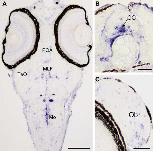
Transgene expression in the central nervous system of zebrafish larvae. (A) At 6 dpf, gfp transcripts expression occurs in the medulla oblongata (Mo) and in scattered cells of the tectum opticum (TeO), preoptic area (POA) and in the medial longitudinal fascicle (MLF). Two symmetric patches of cells located at the boundary between the mesencephalon and the myelencephalon expressed high levels of gfp transcripts (asterisks). (B): At 13 dpf, transgene expression is observed in the central canal (CC) of the spinal cord. (C): At 36 dpf, the transgene is expressed in scattered cells of the olfactory bulb (Ob). EXPRESSION / LABELING:
|
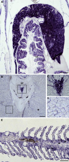
The transgene is expressed in miscellaneous tissues of adult transgenic zebrafish. The gfp transcripts were detected using the whole-mount RNA in situ hybridization with a digoxigenin-labeled antisense riboprobe. (A) gfp transcripts were detected in the hepatocytes of the liver (Li) and in the epithelial cells of the intestine (Int). (B) Transgene expression in the spinal cord (SP) and interstitial cells of the skeletal muscle (M). (C) Inset of the spinal cord. (D) Inset of the skeletal muscle. (E) Note higher transgene expression levels in cells located at the base of the branchial lamellae (black arrows). Scale bars represent 50 μm with the exception of panel B (200 μm). EXPRESSION / LABELING:
|
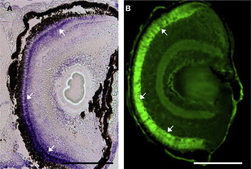
Transgene expression in the retina of hCMV-IE:eGFP transgenic fish. gfp transcript (A) and protein (B) are located in the cone photoreceptor cells (white arrows) of the retina. EXPRESSION / LABELING:
|
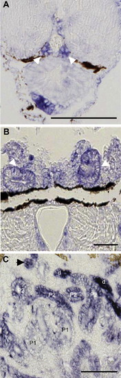
Transgene expression in the kidney of hCMV-IE:eGFP transgenic fish. gfp transcripts were detected in the pronephric ducts and intermediate renal tubules (white arrowheads) of 6 dpf (A) and 13 dpf (B) old larvae, respectively. In adult animals (C), gfp transcripts were observed in the epithelial cells of the renal tubules. Note that transgene expression levels increased from proximal early (P1) to proximal late (P2) and distal (d) tubules. |
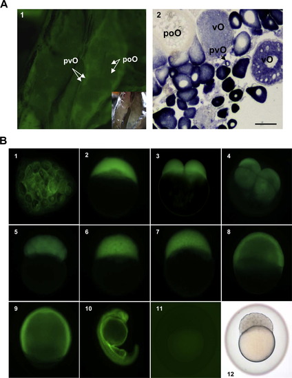
hCMV:eGFP transgene is expressed in ovary. (A) Transgene expression occurs in previtellogenic and vitellogenic oocytes within the ovary. (1) Fluorescence is mainly observed in the primary oocytes; (2) gfp transcripts were detected in the ovary of sexually mature transgenic female adults using the whole-mount RNA in situ hybridization and a digoxigenin-labeled antisense riboprobe. The gfp transcripts were observed in the cytoplasm of previtellogenic (pvO) and vitellogenic (vO) oocytes. No signal was observed in pre-ovulating oocytes (poO). Scale bar represent 150 μm. (B) Maternal inheritance of the GFP protein in the hCMV:eGFP transgenic zebrafish line. (1) The fluorescence is observed in the vitellus of stripped fully mature oocytes before fertilization; (2) one cell stage; (3) 2 cells stage; (4) 4 cells stage; (5) 32 cells stage; (6) 256 cells stage; (7) high; (8) 50% epiboly; (9) 75% epiboly; (10) 24 dpf embryo; (11) zygote at 256 cells stage resulting from the cross of a transgenic male and a wild type female shows no fluorescence; (12) Brightfield image of (11). EXPRESSION / LABELING:
|

Unillustrated author statements EXPRESSION / LABELING:
|
Reprinted from Gene expression patterns : GEP, 13(3-4), Mella-Alvarado, V., Gautier, A., Le Gac, F., and Lareyre, J.J., Tissue and cell-specific transcriptional activity of the human cytomegalovirus immediate early gene promoter (UL123) in zebrafish, 91-103, Copyright (2013) with permission from Elsevier. Full text @ Gene Expr. Patterns