
Fig. 2
- ID
- ZDB-FIG-130510-21
- Publication
- Mella-Alvarado et al., 2013 - Tissue and cell-specific transcriptional activity of the human cytomegalovirus immediate early gene promoter (UL123) in zebrafish
- Other Figures
- All Figure Page
- Back to All Figure Page
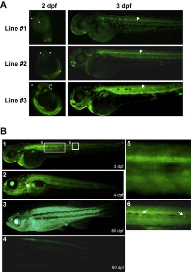
External fluorescence observed in hCMV-IE:eGFP transgenic zebrafish. (A) Transgenic zebrafish were observed with a macroscope equipped with a GFP filter. A similar tissue distribution of transgene expression was observed in three independent transgenic zebrafish lines although different expression levels were observed. Note that transgenic zebrafish lines #1, #2 and #3 showed low, medium and high transgene expression levels, respectively. Transgene was mainly expressed in the neurons of the olfactory placodes (asterisks), the neuromasts, and in the central canal of the spinal cord (arrowheads). (B) Transgene expression pattern during ontogeny: (1) 3 day-old hatched embryo; (2) 4 day-old larvae, the white arrow indicates the liver; (3) 60 day-old transgenic juvenile; (4) 60 day-old wild type (WT) juvenile; (5) inset of 1 showing a high magnification of the spinal cord; (6) inset of 1 showing a high magnification of the lateral neuromasts (white arrows). |
| Gene: | |
|---|---|
| Fish: | |
| Anatomical Terms: | |
| Stage Range: | Long-pec to Days 45-89 |
Reprinted from Gene expression patterns : GEP, 13(3-4), Mella-Alvarado, V., Gautier, A., Le Gac, F., and Lareyre, J.J., Tissue and cell-specific transcriptional activity of the human cytomegalovirus immediate early gene promoter (UL123) in zebrafish, 91-103, Copyright (2013) with permission from Elsevier. Full text @ Gene Expr. Patterns