
- Title
-
Ccm3 functions in a manner distinct from Ccm1 and Ccm2 in a zebrafish model of CCM vascular disease
- Authors
- Yoruk, B., Gillers, B.S., Chi, N.C., and Scott, I.C.
- Source
- Full text @ Dev. Biol.
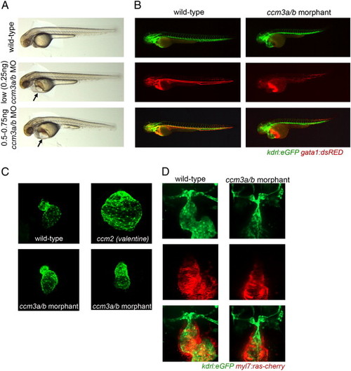
Morpholino knockdown of ccm3a/b in zebrafish embryos causes cardiovascular defects distinct from ccm1/2 mutants. (A) Light images of ccm3a/b morpholino injected embryos at 54 hpf. Morphant embryos display a severe pericardial edema and complete circulatory block, but no gross morphological defects when injected with 0.25–0.75 ng ccm3a/b ATG morpholino. (B) Fluorescent microscopy images of ccm3a/b morpholino injected and non-injected double transgenic kdrl:EGFP; gata1:dsRED embryos at 54 hpf. Morphants lack circulation with blood pooling at the yolk. (C) Confocal images of the ccm3a/b morpholino injected Tg(myl7:EGFP)twu34 wildtype and ccm2 (valentine) mutant embryos. At 54 hpf, ccm3a/b morphant hearts are dysmorphic but not enlarged, unlike the ccm2 (valentine) mutants whose hearts appear grossly dilated. (D) Confocal images of ccm3a/b morpholino injected and non-injected double transgenic kdrl:EGFP, myl7:ras-cherry embryos. Injection of morpholino causes thinning of the ventral aorta and the first aortic arches by 54 hpf. |
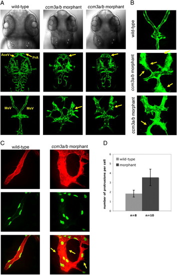
Morpholino knockdown of ccm3a/b causes gross dilation and mispatterning of cranial vessels. (A) At 54 hpf, confocal z-stack projections of dorsal head vasculature showed enlargements in the prosencephalic arteries (PrA), mesencephalic veins (MsV), and anterior cerebral veins (AceV) in embryos injected with 0.75 ng ccm3a/b morpholino (> 90%, n = 62). (B) Morpholino knockdown of ccm3a/b results in greater number of protrusions evident in endothelial cells of the dilated cranial vasculature (yellow arrows). (C) MsVs of the wildtype and ccm3a/b morphant embryos imaged at higher magnification in kdrl:HsHRAS-mCherry; fli1:nlsEGFP transgenic background. (D) ccm3a/b morphant embryos have a significantly higher number of cell protrusions per endothelial cell compared to wildtype (p-value < 0.01, n refers to number of embryos analyzed, with 3–5 cells counted per embryo). |
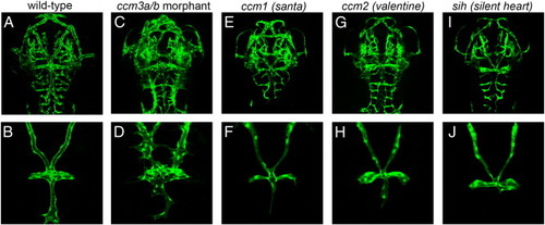
The ccm3a/b cranial vasculature phenotype is specific to ccm3 function and not due to lack of blood flow. Neither santa or valentine mutant embryos display severe vascular dilations or mispatterning seen upon morpholino mediated ccm3a/b knockdown (for san, n = 21, for vtn, n = 32) (E-H) sih embryos similarly displayed no dilations in their cranial vasculature (n = 25) (I and J). Deep confocal z-stack projections (A, C, E, G and I) or z-stacks of dorsal views of the cranial vasculature (B, D, F, H and J) are shown. |

Morpholino knockdown of the GCKIII gene stk25b results in heart and vasculature defects similar to those seen in ccm3a/b morphants. (A) Confocal z-stack projections of dorsal head vasculature of stk25a and stk25b morphants at 54 hpf. Injection of stk25a morpholino (2 ng) caused misconnections in cranial vasculature; however, no appreciable vessel enlargements were observed. Injection of stk25b morpholino (6 ng) caused dilation of prosencephalic arteries (PrA), anterior cerebral veins (AceV) and severe mispatterning of the mesencephalic veins (MsV) in over 75% of the embryos imaged (yellow arrows, n > 80). (B) Light images of stk25a and stk25b morphants. stk25a morphants displayed a pericardial edema, severe yolk extension and trunk elongation defects. The stk25b morphant phenotype was more similar to ccm3a/b morphant morphology, with pericardial and venous plexus edemas. |
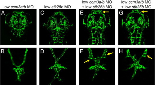
Co-injection of suboptimal ccm3a/b (0.25 ng) and stk25b (3 ng) MO results in cranial vasculature defects at 54 hpf. Embryos injected with low doses of ccm3a/b (0.25 ng) or stk25b morpholino (3 ng) showed no obvious defects in their cranial vessels (A-D). Co-injection of both morpholinos at the same dosages resulted in gross dilation of the prosencephalic arteries (PrA), anterior cerebral veins (AceV), as well as enlargement and mispatterning of the mesencephalic veins (MsV) in 71% of the embryos (E–H, yellow arrows, n = 83). |
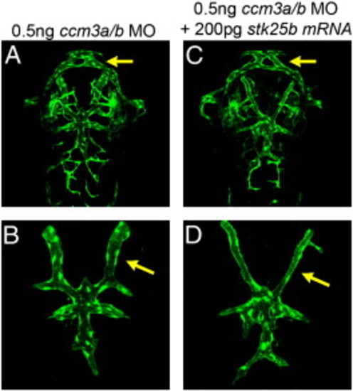
stk25b overexpression in ccm3a/b morphants rescues ccm3a/b cardiovascular phenotype. Confocal z-stack projections of dorsal head vasculature in stk25b RNA (200 pg) injected and non-injected ccm3a/b morphants (0.5 ng ccm3a/b MO). Enlargement of the PrA and AceV in the vasculature were decreased in 68% of ccm3a/b morphants upon injection of stk25b mRNA (A–D, yellow arrows, n = 82). |
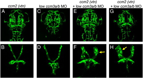
Injection of suboptimal doses of ccm3a/b ATG morpholino into ccm2/vtn mutants results in very severe cranial vascular defects. Gross dilations of the AceV, PrA and MsV vessels were observed upon injection of 0.25 ng ccm3a/b ATG morpholino into ccm2/vtn embryos (A–H, yellow arrows). |
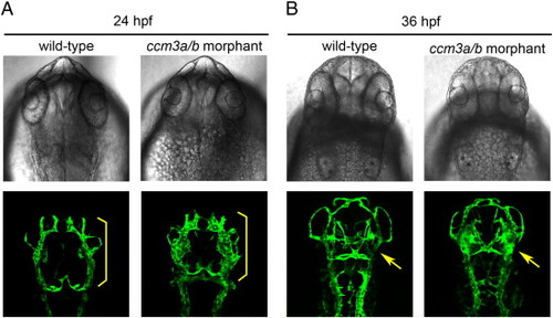
Cranial vasculature defects can be observed in ccm3a/b morphants at 24 and 36 hpf. At 24hpf, ccm3a/b morphants (0.75 ng ccm3a/b MO) display enlarged primordial midbrain channels (PMCs), compared to wildtype embryos (yellow brackets). At 36hpf, morphant midcerebral veins, which sprout from the PMC, also appeared enlarged compared to wildtype (yellow arrows). |
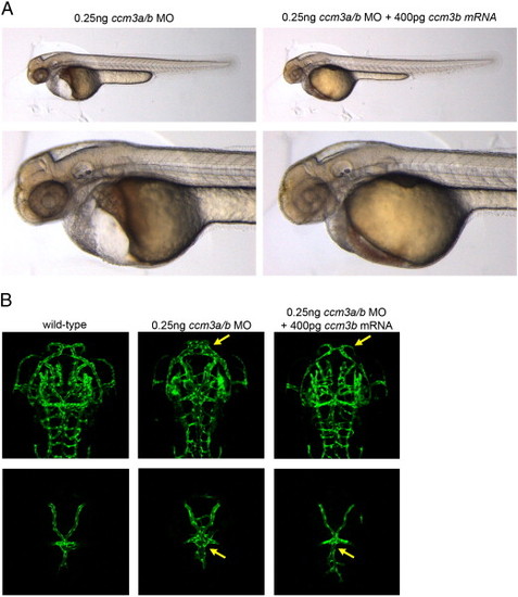
Cardiovascular defects caused by morpholino-knockdown of ccm3a/b can be rescued by overexpression of ccm3b mRNA. (A) Light images of ccm3b mRNA injected and non-injected ccm3a/b morphant embryos at 54 hpf. The pronounced pericardial edema and circulatory block observed in 93% of the embryos upon 0.25 ng ccm3a/b morpholino injection (n = 83) are rescued by co-injection of 400 pg ccm3b mRNA. 63% of ccm3a/b morphant embryos injected with ccm3b mRNA had normal heart morphology and completely restored circulation (n = 76). (B) Confocal z-stack projections of dorsal head vasculature in ccm3a/b morphants indicate a decrease in the dilation and misconnections in cranial vessels upon ccm3b mRNA injection (yellow arrows). |
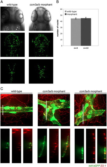
Cranial vasculature enlargements seen in ccm3a/b morphants is not due to overproliferation of endothelial cells in the affected vessels. (A) Confocal z-stack projections of dorsal head vasculature in wildtype and ccm3a/b morphant embryos in the Tg(fli1:nlsEGFP) background at 54 hpf. (B) Endothelial cell nuclei were quantified in ccm3a/b morpholino injected (0.75 ng) and non-injected mesencephalic veins using the Tg(fli1:nlsEGFP) line. There was no statistically significant difference between the endothelial cell numbers in morphants compared to wildtype (p-value = 0.78). (C) Tg(kdrl:EGFP) embryos at 54 hpf were stained with mouse anti-ZO-1, and confocal images were analyzed for ZO-1 expression (red), and blood vessels (green). Cross-sections of the z-stack projections, as indicated by the white bars, revealed that wildtype mesencephalic veins (MsVs) were properly lumenized with strong ZO-1 expression at tight junctions along the circumference of the vessels. ccm3a/b morphant MsVs were enlarged, and in many instances failed to lumenize. ZO-1 expression in the morphants appeared disorganized and showed decreased co-localization with the vasculature (green). |
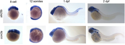
Similarity of stk25b and ccm3a expression patterns. Both stk25b and ccm3a were found to be expressed maternally at the 8 cell-stage. Both genes displayed a broad expression profile at the 12 somite stage (15hpf). At 24 hpf, their expression became more restricted to the head region and remained so at 48 hpf. |

Unillustrated author statements |
Reprinted from Developmental Biology, 362(2), Yoruk, B., Gillers, B.S., Chi, N.C., and Scott, I.C., Ccm3 functions in a manner distinct from Ccm1 and Ccm2 in a zebrafish model of CCM vascular disease, 121-131, Copyright (2012) with permission from Elsevier. Full text @ Dev. Biol.