
- Title
-
Jamb and jamc are essential for vertebrate myocyte fusion
- Authors
- Powell, G.T., and Wright, G.J.
- Source
- Full text @ PLoS Biol.

jamb and jamc are co-expressed in myoblasts. (A–B) Wholemount in situ hybridisation of jamb (A) and jamc (B) show expression in myoblasts during somitogenesis (10–13 somites, 17–18 somites, and 21 somites). Expression is attenuated in the myotome after the completion of primary myogenesis (24 h. p. f.) and then later upregulated in craniofacial (cf), pectoral fin (arrows), and hypaxial (open arrowheads) muscle mesoderm (48 h. p. f.). Scale bars represent 50 μm. EXPRESSION / LABELING:
|

jamb and jamc are essential for myocyte fusion in vivo. (A) Schematics of Jamb and Jamc extracellular proteins. Red stars denote sites of mutation in HU3319 and sa0037 alleles. (B–C) Confocal microscopy images of fast-twitch muscle in wild-type, jambHU3319, and jamcsa0037 48 h. p. f. (B) and 120 h. p. f. (C) Embryos labelled with membrane targeted RFP (mRFP, cyan; B) or phalloidin-Alexa488 (cyan; C) and DAPI (red) show overabundant, mononuclear myofibres in both mutants. (D) Confocal microscopy images of uninjected, jamb, and jamc translation-blocking morpholino-injected wild-type embryos, stained with DAPI (red) and phalloidin-Alexa488 (cyan) to stain F-actin in fast muscle fibres. The morpholino-injected embryos replicate the jam mutants′ phenotype. Myotomes 12–13 shown, anterior left. Scale bars represent 50 μm. |

Fast and slow-twitch muscle fibres are correctly specified in jambHU3319 and jamcsa0037 embryos. (A) Superimposed confocal microscopy images of 24 h. p. f. wild-type, jambHU3319, and jamcsa0037 embryos stained with monoclonal antibody F59, which detects slow-muscle-specific myosin heavy chain (sMyHC), show normal slow muscle development in both mutants. (B) Single optical sections of 48 h. p. f. wild-type, jambHU3319, and jamcsa0037 embryos stained with monoclonal antibody EB165, which detects fast-muscle-specific myosin heavy chain (fMyHC), show that the medial mononuclear fibres in both mutants have differentiated as fast-twitch muscle. Myotomes 12–13 shown, anterior left. Scale bars represent 50 μm. |
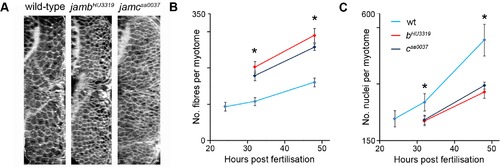
Fast muscle fibres are supernumary in jambHU3319 and jamcsa0037 embryos. (A) Transverse sections projected from confocal microscopy images of 48 h. p. f. embryos labelled with mRFP. (B–C) Graphs showing a significant overabundance (1.6–1.8-fold) of muscle fibres in mutants compared to wild-type (B) and calculated muscle nuclei number in wild-type and mutant embryos between 24 and 48 h. p. f. (C). Asterisk denotes pd0.001; one-tailed Student′s t test; error bars represent standard deviation, see Tables S1 and S2 for number of embryos in each sample. PHENOTYPE:
|
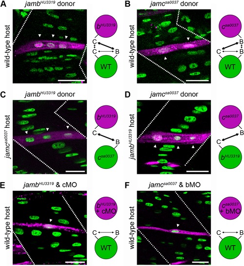
Interaction between jamb and jamc is necessary and required in trans for myocyte fusion. (A–D) Fluorescent dextran-labelled cells (magenta) from jambHU3319 and jamcsa0037 donors can form multinucleate fibres with wild-type (A, B), jamcsa0037 (C), or jambHU3319 (D) host cells, respectively. (E, F) Transplanted cells from doubly-deficient donors fail to fuse with wild-type host cells, suggesting both proteins are required and interact in trans. Confocal microscopy images from 48 h. p. f. embryos; anterior left. Schematics illustrate potential binding of Jamb and Jamc between donor (magenta) and host (green) cells in each experiment. bMO, cMO indicate jamb or jamc translation-blocking morpholino-injected donor embryos. Dotted lines indicate the position of myotome boundaries; arrowheads indicate nuclei within labelled fibres. Nuclei stained with DAPI (green). Scale bars represent 20 μm. |
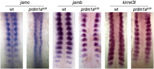
jamc expression is regulated by prdm1a. Wholemount in situ hybridisation against jamc (left panels), jamb (middle panels), and kirrel3l (right panels) shows that jamc is ectopically expressed in premigratory slow muscle precursors (adaxial cells) of prdm1atp39 mutants that later inappropriately fuse to fast muscle myocytes. In contrast, jamb and kirrel3l are expressed in fast muscle myoblasts in both wild-type and prdm1atp39 mutant embryos. Flatmounted embryos at 10–13 somite stage; anterior top. EXPRESSION / LABELING:
|
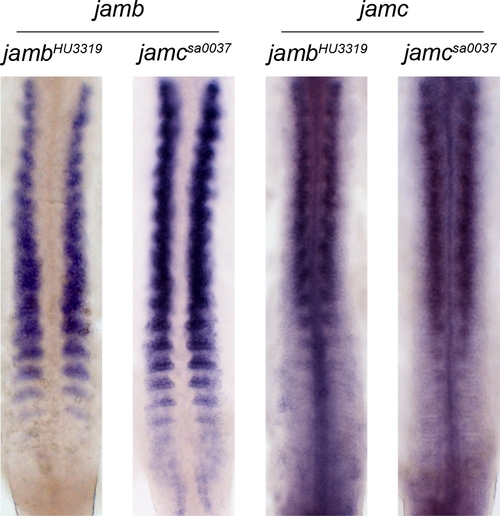
Expression of jamb and jamc in jambHU3319 and jamcsa0037 mutant embryos. In situ hybridisation of jamb (left two panels) and jamc (right two panels) riboprobes to jambHU3319 and jamcsa0037 embryos at 17–18 somites stage; anterior top. Both genes are expressed in fast muscle myoblasts in both jambHU3319 and jamcsa0037 mutants as observed in wild-type embryos. EXPRESSION / LABELING:
|

Combined knockdown of jamb and jamc does not result in a synthetic myogenesis phenotype. (A, B) Antisense morpholino oligonucleotide knockdown of expression of jamc in jambHU3319 embryos (A, right) or jamb in jamcsa0037 embryos (B, right) does not result in any further disruption of myogenesis than that observed in jambHU3319 (A, left) or jamcsa0037 (B, left) at 48 h. p. f., suggesting no synthetic effect of combined knockdown of both genes. Single confocal microscopy images of myotomes 12–13 in 48 h. p. f. embryos, stained for F-actin (cyan) and nuclei (red). Anterior left; scale bars represent 50 μm. PHENOTYPE:
|
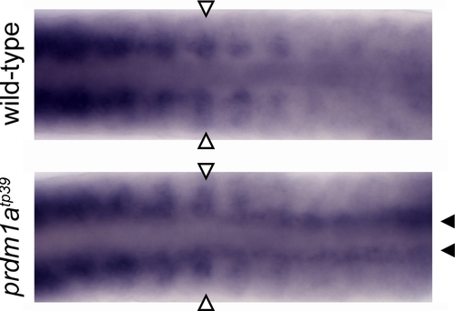
jamc expression in fast muscle myoblasts and adaxial cells of prdm1atp39 mutants. jamc is expressed in fast muscle myoblasts (open arrowheads) and ectopically expressed in premigratory slow muscle precursors (adaxial cells; closed arrowheads) of prdm1atp39 mutant embryos (bottom). Flatmounted wild-type sibling (top) and prdm1atp39 mutant embryos (bottom) at 18–20 somites stage, hybridised to jamc; anterior left. EXPRESSION / LABELING:
|