
- Title
-
Cell adhesion molecule cadherin-6 function in zebrafish cranial and lateral line ganglia development
- Authors
- Liu, Q., Dalman, M.R., Sarmah, S., Chen, S., Chen, Y., Hurlbut, A.K., Spencer, M.A., Pancoe, L., and Marrs, J.A.
- Source
- Full text @ Dev. Dyn.

cdh6 expression in the cranial and lateral line ganglia of embryonic zebrafish. All panels show lateral views of the hindbrain region of whole-mount embryos (anterior to the left and dorsal up) processed for in situ hybridization using a cdh6 cRNA probe. Panel C is a higher magnification of the post otic area showing the newly formed primordium of the post lateral line (pPLL, dashed line indicating the boundary of its posterior 2/3). The arrow points to one of a few cdh6-expressing cells in the pPLL. a, anterior lateral line placode area; c, cerebellum; gAd, anterodorsal lateral line ganglion; gAv, anteroventral lateral line ganglion; gM/X, medial lateral line and vagal ganglia; gP, posterior lateral line ganglion; gV, trigeminal ganglion; h, hindbrain; op, otic placode; ov, otic vesicle (indicated by the dashed line); PP, postotic placode; sag, statoacoustic ganglion. Scale bar = 50 μm. EXPRESSION / LABELING:
|
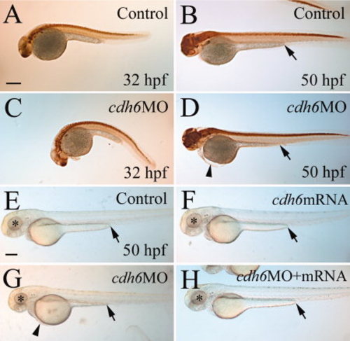
Overall cdh6 loss-of-function phenotype. Lateral views (anterior to the left and dorsal up) of whole-mount embryos processed for anti-HuC/D immunoperoxidase staining. A–D: The morphants (C,D, injected with cdh6 MO2 [morpholino oligonucleotide 2] showing moderate phenotype) were similar in body and yolk size as uninjected control embryos (A,B), but had smaller head and eyes, curved body at younger (e.g., 32 hours postfertilization) stages, shortened yolk extension and edema in the thorax, as shown in our previous publication (Liu et al., 2008a). E–H: Live embryos raised in PTU (1-phenyl-2-thiourea, 0.003%) -treated fish water. The eyes are indicated by asterisks, edema in the thorax is indicated by an arrowhead, and the end of the yolk extension is indicated by an arrow. Scale bar = 250 μm for all panels. |

Development of the cranial and lateral line ganglia requires Cdh6 function. A–J: Anti-HuC/D immunoperoxidase staining of embryos, showing lateral views (anterior to the left and dorsal up) of the head region. K,L: Histograms representing the area/size (square microns; n = 13 for all measured ganglia) of anti-HuC/D labeled cranial and lateral line ganglia, comparing control embryos (gray bars), cdh6 morphants (dark bars) and cdh6 mRNA rescued embryos (bars with diagonal lines). One asterisk indicates significant difference (P = 0.0013), whereas three asterisks indicate highly significant difference (P < 0.001). gX, vagal ganglion; ot, optic tectum. Other abbreviations are the same as in Figure 1. Scale bars = 100 µm in A,F, 50 μm in B–E,G–L. |
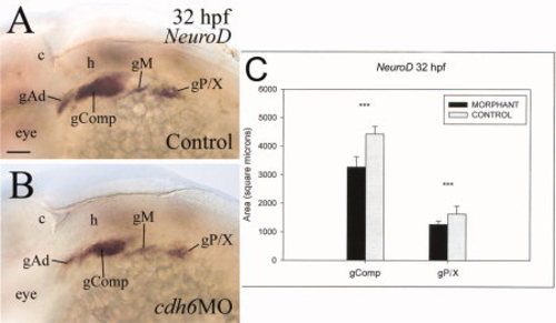
Cdh6 function is required for differentiation of NeuroD-positive cranial and lateral line ganglia. A,B: Lateral views (anterior to the left and dorsal up) of the hindbrain region of whole-mount embryos processed for in situ hybridization using the NeuroD cRNA probe. C: Comparisons in area size of two NeuroD-labeled areas between control (gray bars) and cdh6 morphants (dark bars). gComp, complex “ganglion” including the facial ganglion, anteroventral lateral line ganglion and statoacoustic ganglion; gM, middle lateral line ganglion; gP/X, posterior lateral line ganglion and precursors of vagal ganglion. Other abbreviations are the same as Figure 1. Scale bar = 50 μm. |

Cdh6 function is required for differentiation of zn-5–positive cranial and lateral line ganglia. All panels are lateral views (anterior to the left and dorsal up) of the head region of whole-mount embryos. Abbreviations are the same as in Figure 1. All images have the same magnification. Scale bar = 50 μm. |
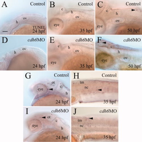
Apoptosis analysis using TUNEL (terminal deoxynucleotidyl transferase–mediated deoxyuridinetriphosphate nick end-labeling) staining. All panels show lateral views of whole-mount embryos (anterior to the left and dorsal up) processed for TUNEL staining. The mid- and hindbrain region focusing on the otic vesicle (A–G,I), while panels H and J are from the body trunk region (H,J). Arrowheads point to some TUNEL-positive cells. nc, notochord; tm, trunk muscles. Other abbreviations are the same as in Figure 1. Scale bar = 50 μm for all panels. |
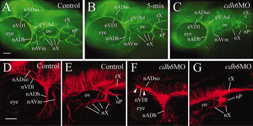
cdh6 loss-of-function defects in cranial and lateral line nerves, demonstrated by anti-acetylated tubulin immunofluorescent staining, in a control embryo (A), an embryo injected with the 5-mismatched MO (B), and a cdh6 morphant (C). The images are lateral views (anterior to the left and dorsal up) of the head region of the whole-mount embryos. D–G: Laser scanning confocal microscopy image projections confirmed results using wide field microscopy: anterior cranial nerves (D,F) and posterior cranial nerves (E,G) in control (D,E) and cdh6 morphant (F,G) embryos. Arrowheads in F indicate two cell clusters of the fragmented gV/Ad. The morphant vagus root (rX, G) was difficult to discern because it was closely opposed to anti-acetylated tubulin-positive brain cells and fiber tracks. Other abbreviations: nADb, buccal ramus of the anterodorsal lateral line nerve; nADso, superior ophthalmic ramus of the anterodorsal lateral line nerve; nAVm, mandibular ramus of the anteroventral lateral line nerve; nVDl, dorsolateral nerve of the trigeminal ganglion; nP, posterior lateral line nerve; nX, vagus nerves; ov, otic vesicle. All images have the same magnification. Scale bars = 50 μm. PHENOTYPE:
|

Normal development of the posterior lateral line nerve in cdh6 morphants. All panels show lateral views of whole-mount embryos (anterior to the left and dorsal up) processed for anti-acetylated tubuline immunoperoxidase staining. Arrows point to the posterior lateral line nerve, while the asterisk indicates the terminus of the nerve. C and D are higher magnifications (same magnification) of the tail region of the embryos in A and B (same magnification), respectively. Scale bar = 200 μm in A,B, 100 μm in C,D. |