
- Title
-
Potential targets of transforming growth factor-beta1 during inhibition of oocyte maturation in zebrafish
- Authors
- Kohli, G., Clelland, E., and Peng, C.
- Source
- Full text @ Reprod. Biol. Endocrinol.

Validation of semi-quantitative RT-PCR for GAPDH (A), 20βHSD (B), FSHR (C), LHR (D), mPR-α (E) and mPR-β (F). PCRs were performed using zebrafish ovarian cDNA as the template, amplified for varying cycle numbers and the density of the PCR products was quantified. Each value represents the mean ± SEM of three replicates in one representative RT-PCR. Representative ethidium bromide stained gel pictures were included. C = negative control; number on each lane represents the number of PCR cycles performed. |

TGF-β1 inhibits mRNA expression of 20β-HSD. (A) Follicles were treated with control medium or 10 ng/ml of TGF-β1 for 2, 6, 12 and 24 hours. (B) Follicles were treated with different concentrations (0, 0.1,1 and 10 ng/ml) of TGF-β1 for 18 hours. (C) Follicles were treated with control medium, hCG (100 IU/ml), TGF-β1 (10 ng/ml), or hCG+ TGF-β1 for 18 hours. Total RNA was extracted and subjected to RT-PCR using primers for 20β-HSD and GAPDH. Each value represents the mean ± SEM of three replicates in one representative RT-PCR reaction. 20β-HSD mRNA levels are expressed as percent of control after normalized with the GAPHD levels. Different letters above the bars denote statistical significance (P < 0.05). *, P < 0.05 vs. control. The insets show representative ethidium bromide stained gels. GAPDH gels are the same for Figs. 4-7. EXPRESSION / LABELING:
|

TGF-β1 stimulates FSHR mRNA expression. (A) Follicles were incubated with control medium or 10 ng/ml of TGF-β1 for 2, 6, 12 and 24 hours. (B) Follicles were treated with different concentrations (0, 0.1,1 and 10 ng/ml) of TGF-β1 for 18 hours. (C) Follicles were treated with medium (control), hCG (100 IU/ml), TGF-β1 (10 ng/ml), or a combination of hCG and TGF-β1 for 18 hours. At the end of each incubation, total RNA was extracted and reverse transcribed. PCR was carried out using primers for FSHR and GAPDH. Each value represents the mean ± SEM of three replicates in one representative RT-PCR. Statistical significance (P < 0.05) is indicated by either an * or a different letter. The insets show the representative ethidium bromide stained gels. EXPRESSION / LABELING:
|
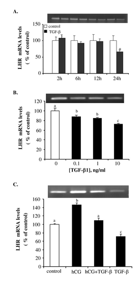
TGF-β1 suppresses LHR mRNA expression. Follicles were treated with (A) 10 ng/ml of TGF-β1 for 2, 6, 12 and 24 hours; (B) 0.1, 1, or 10 ng/ml of TGF-β1 for 18 hours; and (C) hCG (100 IU/ml), TGF-β1 (10 ng/ml), either alone or in combination, for 18 hours. Each value represents the mean ± SEM of three replicates in one representative RT-PCR. Different letters denote statistical significance (P < 0.05). The insets show the original ethidium bromide stained gels. EXPRESSION / LABELING:
|

TGF-β1 has no effect on mPR-α mRNA expression. Follicles were incubated with (A) 10 ng/ml of TGF-β1 for 2, 6, 12 and 24 hours; (B) different concentrations of TGF-β1 for 18 hours; and (C) medium only (control), hCG (100 IU/ml), TGF-β1 (10 ng/ml) or hCG + TGF-β1 for 18 hours. Each value represents the mean ± SEM of three replicates in one representative RT-PCR. The insets show representative ethidium bromide stained gels. Neither hCG nor TGF-β1 had an effect on mPR-α mRNA expression. EXPRESSION / LABELING:
|
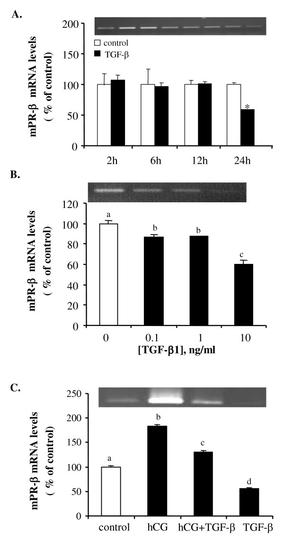
TGF-β1 downregulates mPR-β mRNA expression. (A) Time course study of the effect of TGF-β1 on mPR-β mRNA expression. Follicles were treated with 10 ng/ml of TGF-β1 for 2, 6, 12 and 24 hours. (B) Follicles were treated with different concentrations (0, 0.1,1 and 10 ng/ml) of TGF-β1 for 18 hours. (C) Follicles were incubated with medium (control), hCG (100 IU/ml), TGF-β1 (10 ng/ml) or hCG + TGF-β1 for 18 hours. Each value represents the mean ± SEM of three replicates in one representative RT-PCR. Different letters or "*" denote statistical significance (P < 0.05). The insets show the representative ethidium bromide stained gels. EXPRESSION / LABELING:
|