
- Title
-
Expression of collapsin response mediator proteins in the nervous system of embryonic zebrafish
- Authors
- Schweitzer, J., Becker, C.G., Schachner, M., and Becker, T.
- Source
- Full text @ Gene Expr. Patterns

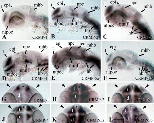
Expression of CRMP mRNAs at 16 hpf. Side views of heads (A–F) or tails (G–L) of whole-mounted embryos with the yolk sac removed are shown (rostral is left, dorsal is up). Probes used are indicated in the individual panels. (A–F) In the head, different CNS regions express CRMP-2, -3, -4 and -5a mRNAs. Presumptive structures showing an in situ hybridization signal are indicated (t=telencephalon, ntpoc=nucleus of the post-optic commissure, epi=epiphysis, npc=area close to the nucleus of the posterior commissure, nmlf=nucleus of the medial longitudinal fascicle, tg=trigeminal ganglion, hb=clusters of cells in the hindbrain). (G–L) In the tail region, expression of CRMP-2, -3, -4 and -5a mRNAs is found in the spinal cord. CRMP-2 and -3 mRNAs are prominently expressed in dorsally located neurons (arrowheads in H,I). Expression of CRMP-4 and -5a mRNAs extends to the caudal tip of the spinal cord (arrowheads in J,K), but labeling intensity is higher towards the rostral, more mature spinal cord (arrows in J,K). CRMP-1 and -5b mRNAs are not detectable at 16 hpf. Scale bar in L=100 μm for A–L. EXPRESSION / LABELING:
|
Expression of CRMP mRNAs in the head of 24 hpf embryos. Side views (A–F) or top views (G–L) of whole-mounted embryos with the yolk sac removed are shown. Probes used are indicated in the individual panels. (A–F) (rostral is left, dorsal is up) CNS regions showing expression of the CRMP mRNAs are indicated in the photomicrographs, with the exception of the midbrain–hindbrain boundary (mhb) that is always indicated for orientation, but shows a strong signal only for CRMP-2, -3 and -5a (tec=tectum mesencephali; all other abbreviations as in Fig. 3). (G–L) (rostral is up) Olfactory placodes (always indicated by arrowheads) express CRMP-2, -3, -4 and -5a mRNAs. Telencephalon (t) and diencephalon (d) express all CRMP mRNAs, but not in the proliferating ventricular zone (asterisks). The eyes, which are already lightly pigmented at 24 hpf, are negative for all CRMP mRNAs. Scale bar in F=100 μm for A–F; scale bar in L=100 mm for G–L. EXPRESSION / LABELING:
|

Differential expression of CRMP mRNAs in the hindbrain and trunk region at 24 hpf. Probes used are indicated in the photomicrographs. (A) In a top view of an embryo labeled with a CRMP-4 probe (rostral is left), serially repeated clusters of neurons (arrowheads) can be observed in the hindbrain (hb). Additional signal is present in the nucleus of the medial longitudinal fascicle (nmlf), the trigeminal ganglion (tg), the acoustic/anterior lateral line ganglion (ac/allg) and the posterior lateral line ganglion (pllg). The otic vesicle (ov) is indicated for orientation. (B,C) In cross-sections of spinal cord (dorsal is up) labeled with CRMP-3 (B) or CRMP-4 (C) cRNA probes, a signal in the lateral domain, containing differentiated neurons, is obvious. Large Rohon-Beard neurons (RhB) strongly express CRMP-4 mRNA. (D–I) Side views of mid-trunk regions are shown (rostral is left, dorsal is up). Pairs of horizontal bars delineate the dorso-ventral extent of the spinal cord. CRMP-2 and -4 mRNAs are prominently expressed in Rohon-Beard neurons, some of which are indicated by arrowheads in E,G. Arrowheads in F indicate CRMP-3 mRNA positive spinal interneurons. Expression of CRMP-5b mRNA is weak in the spinal cord (arrowheads in I) and CRMP-1 mRNA was not detected in the spinal cord. Scale bar in A, 100 μm; scale bar in C, 25 μm for B, C; scale bar in I, 100 μm for D–I. EXPRESSION / LABELING:
|

Unillustrated author statements EXPRESSION / LABELING:
|
Reprinted from Gene expression patterns : GEP, 5(6), Schweitzer, J., Becker, C.G., Schachner, M., and Becker, T., Expression of collapsin response mediator proteins in the nervous system of embryonic zebrafish, 809-816, Copyright (2005) with permission from Elsevier. Full text @ Gene Expr. Patterns