
- Title
-
Overexpression of the forebrain-specific homeobox gene six3 induces rostral forebrain enlargement in zebrafish
- Authors
- Kobayashi, M., Toyama, R., Takeda, H., Dawid, I.B., and Kawakami, K.
- Source
- Full text @ Development

Expression of six3 in early embryogenesis. (A-C,I) Lateral views with dorsal to the right; (D-H), dorsoanterior views with posterior to the top. (A) 55% epiboly; six3 starts being expressed in the hypoblast of the embryonic shield. (B) 75% epiboly; six3 signal is localized in the anterior axial mesendoderm. (C) 80% epiboly; embryo was hybridized with six3 and ntl probes together using the same color; six3 is expressed only in the anterior (black triangle), whereas ntl is expressed posteriorly (open triangle). (D-H) Tail bud stage; embryos were hybridized with six3 (D), gsc (E), six3+gsc (F), six3+ntl (G), six3+pax2 (H), using the same color. (D-F) six3 is expressed in ectodermal cells of the prospective forebrain (black triangle). six3 expression in the anterior axial mesendoderm is delimited to the polster (arrow), while gsc expression is posteriorly expanded (open triangle). (G) six3 is not expressed in the presumptive notochord; there is a gap between six3 (black triangle) and ntl (open triangle) mRNA-expressing cells. (H) six3 is expressed in the prospective forebrain, indicated by a gap between six3 (black triangle) and pax2 (open triangle) mRNA-expressing cells. (I) Twosomite stage; six3 expression is sharply delimited to the rostral brain. Scale bar, 200 μm. |

six3 expression in the head ectoderm. Dorsoanterior views (A-C, posterior to the top) and corresponding transverse sections (D-F). Dotted lines in A-C indicate approximate levels of the sections. (A,D) Two-somite stage; six3 is expressed in both ectodermal and mesendodermal cells. (B,E) Six-somite stage; six3 mRNA is present primarily in ectodermal cells. (C,F) 16-somite stage; six3 expression is restricted to the eye and the rostral regions of the forebrain. The section shows that six3 expression in both retina and lens is dominant in surface cells, while the neural tube is largely six3 negative. Abbreviations: ec, ectoderm; l, lens; me, mesendoderm; nr, neural rod; nt, neural tube; r, retina. Scale bars, 100 μm. EXPRESSION / LABELING:
|

six3 expression in later development. (A) Lateral view of 24- hour embryo showing six3 expression at the rostral surface of the forebrain (bracket); the eyes were removed. (B,C) Lateral (B) or frontal (C) view of six3 expression at 36 hours. The six3 signal (blue) is localized in the medial telencephalon (arrow), optic stalk (open triangle) and near the olfactory nerve (thin arrow). Axons were stained with antibody against acetylated α-tubulin (brown). (D,E) Lateral views of six3 (D) or lim3 (E) expression at 48 hours. six3 is expressed in the pituitary anlage (black triangle). Additional abbreviations: ac, anterior commissure; cb, cerebellum; dd, dorsal diencephalon; e, epiphysis; hy, hypothalamus; poc, postoptic commissure; rh, rhombomeres; t, telencephalon; te, tectum. Scale bars, 200 μm. |
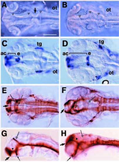
Disorganized head formation in six3 mRNA-injected zebrafish embryos. 20 pg of globin (A,C,E,G) or six3 mRNA (B,D,F,H) were injected into 2-cell-stage zebrafish embryos; see Fig. 7 for quantitative results. (A,B) Dorsal views at 24 hours. Three ventricles (open triangles) in the brain are filled with masses of cells and the midbrain-hindbrain boundary (arrow) is disorganized in six3 mRNA-injected embryos. (C,D) Dorsal views of Islet-1 expression at 24 hours. The region between AC and epiphysis (bracket) is enlarged in six3 mRNA-injected embryos. Likewise, the distance between left and right trigeminal ganglia is expanded. (E-H) Dorsal (E,F) or lateral views (G,H) of 36 hour embryos labeled with antibody against acetylated α-tubulin. AC (arrow), POC (thin arrow), SOT (black triangle) and region surrounded by them are enlarged, while axons in PC (open arrow) are reduced in six3 mRNA-injected embryos. Additional abbreviations: ot, otic vesicle; tg, trigeminal ganglion. Scale bar, 200 μm. |

The dorsal neural tube expands in six3 mRNA-injected embryos. Transverse sections of embryos injected with globin (A,C) or six3 mRNA (B,D). (A,B) Six-somite stage; sections through the prospective forebrain and optical vesicles. Dorsal (bracket) but not ventral region contains extra cells in six3 mRNA-injected embryos. The size of cells is basically unchanged. (C,D) 24 hours; Sections through the midbrain. Structure of dorsal portion of the neural tube is disorganized, but cell number is not changed substantially. Scale bar, 100 μm. |

Effects of six3 overexpression on expression patterns of brain subregion-specific genes. Globin (A,C,E,G) or six3 mRNA-injected (B,D,F,H) embryos. (A,B) Lateral views of lim5 expression at the 16- somite stage. The rostral lim5-negative region (bracket) expands in six3 mRNA-injected embryos. The apparent posterior displacement of ventral lim5-positive cells (arrows) is smaller than that of dorsal cells. (C,D) Lateral views of otx2 expression at 29 hours. The rostral otx2-negative region (bracket) is enlarged in six3 mRNA-injected embryos, while the otx2 mRNA-expressing region moves posteriorly and becomes shorter. (E,F) Lateral views of shh expression at 24 hours; shh expression is not changed substantially. (G,H) Lateral views of emx2 expression at 24 hours. The emx2-positive region in the telencephalon, but not in the hypothalamus, is enlarged. Scale bar, 200 μm. |

Accumulation of pax2-positive cells in six3 mRNA-injected embryos. pax2 expression in globin (A,C,E,G) or six3 (B,D,F,H) mRNA-injected embryos was analyzed at the 10-somite stage (A,B) or at 24 hours (C-H). (A,B) pax2 mRNA-expressing cells in the anlage of the optic vesicles are increased in six3 mRNA-injected embryos, while expression at the prospective midbrain-hindbrain boundary and at the otic placode is unchanged. (C,D) In control embryos pax2-positive optic stalk cells (arrow) are located in the ventral diencephalon, while these cells are located more dorsally in six3 mRNA-injected embryos. (E,F) In the eye, the pax2-positive region at the choroid fissure, i.e., the base of the optic stalk (arrow), is enlarged in six3 mRNA-injected embryos. Since the lens is unaffected, a gap arose between lens and retina (thin arrow). (G,H) Oblique ventral-lateral views of the optic stalk and eyes show that the base of optic stalk is substantially enlarged in six3 mRNA injected embryos. Schematic views show six3 expression as blue areas. Circles drawn in solid and dotted lines indicate the proximal and distal eyes, respectively. Scale bars, 100 μm. Additional abbreviations: l′, lens in the distal eye; mh, midbrain-hindbrain boundary; r′, retina in the distal eye. |