
- Title
-
Organization of hindbrain segments in the zebrafish embryo
- Authors
- Trevarrow, B., Marks, D.L., and Kimmel, C.B.
- Source
- Full text @ Neuron
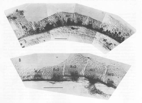
Antibodies Reveal the Segmental Organization of the Embryonic Zebrafish Hindbrain (A) Labeling by the zn-1 antibody, which recognizes many or all neurons (Table 1). A cluster of labeled cells occupies the center of each neuromere, and much smaller groups of positive cells are beginning to develop in the border regions between some of the neuromeres. The hindbrain-midbrain (cerebellar region: cb) boundary is shown to the left. The fourth ventricle is dorsal to most of the hindbrain tissue. (B) Labeling by the zn-12 antibody, which at early stages recognizes a subset of neurons present in the segment centers. Sagittal sections at 24 h are shown rostral to the left and dorsal up. All subsequent figures showing sagittal views are oriented the same way. Here and throughout the paper, naming of the hindbrain segments follows previous conventions (Metcalfe et al., 1986; Hanneman et al. 1988); there are three rostral segments, Ro1-Ro3, three middle ones, Mi1-Mi3, and three caudal divisions, Ca1-Ca3. Scale bars: 50 μm. |
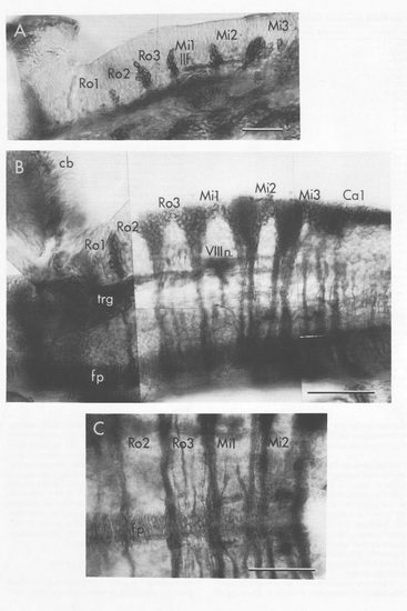
The Zn-5 Antibody Labels Clusters of Neurons and Their Commissural Axons That Develop Near the Segment Borders (A) Sagittal section at 29 h. The clusters are located dorso-lateral to the lateral longitudinal fasciculus (llf, a predominantly sensory pathway that includes descending fibers of the trigeminal nerve; see P.Z. Myers, W.K. Metcalfe, and C.B. Kimmel, submitted), revealing their location in the alar plate. The developing pharyngeal arches ventral to the hindbrain and the otic vesicle (otv) are also labeled. (B and C) Views of the hindbrain in 48h embryos dissected after fixation. (B) Dorsolateral oblique view. The labeled cell clusters lie in the border regions dorsal (aand lateral) to the eighth nerve fibers (VIIIn.), which enter the brain in the Mi1 segment. Note that the neuronal clusters in the Mi1-2, Mi2-3, and Mi3-Ca1 border regions project two large bundles of commissural axons, as contrasted with the clusters located more rostrally, which project only a single large bundle. The floor plate cells (fp) at the ventral midline form a continuous, nonsegmented row in the hindbrain (and also in the midbrain; data not shown). cb: cerebellum, trg:trigeminal ganglion. (C) Dorsal view of the midline region. The double commissures in (B) and (C) are in the same locations. In addtion, one or two labeled commissures extend across the centers of some segments (e.g. Ro3). Scale bars: 50 μm. |

The FRet 8 Antibody Labels a Ladder-like Array of Axons in a Horizontal Section through the Hindbrain and Midbrain at 48 h. Labeled commissures cross near the segment borders (arrow-heads). The commissures are out of the plane of section where they cross the midline rostral (to the top) to Mi3. Many (seven or more) discrete longitudinal fiber pathways are visible bilaterally in the Ro2-Mi2 segments. There is a similar number of longitudinal fascicles in the midbrain (mbr) near the nucleus of the medial longitudinal fascicle (nuc mlf). Faintly labeled lateral clusters of neurons (asterisks) are visible between the Mi1 and Mi2 segments. The posterior lateral line ganglion (pllg) is caudal to the Ca1 segment. Scale bar: 50 μm. EXPRESSION / LABELING:
|

Neuropil, Labeled by Znp Antibodies, First Develops in the Hindbrain Segment Centers (A) Labeling by znp-1 is localized in the neuromuscular junction region of the extrinsic eye muscles, suggesting it recognizes a synaptic component. The other znp antibodies label muscle similarly and also show punctate staining at the bases of hair cells, where synapses have been identified (Metcalfe et al., 1985). (B and C) Sagittal sections of the hindbrain at 48 hr. In (B), neuropil present in the segment centers is labeled in a coarse punctate fashion by znp-4. In (C), the znp-1 antibody labels the same region, but in a more delicate fashion. Longitudinal tracts are also labeled as well as a thin dorso-ventrally aligned region occasionally visible near the segment borders (arrowheads). Scale bars: 25 μm. EXPRESSION / LABELING:
|
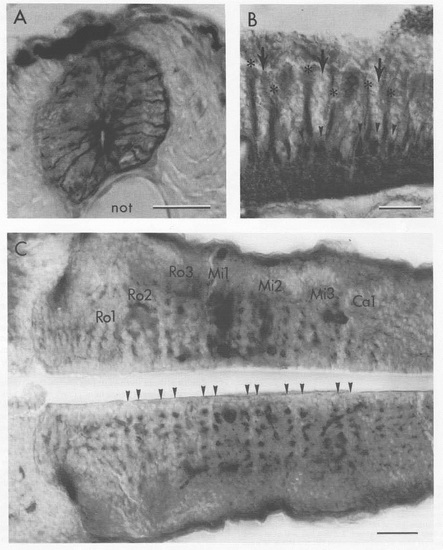
Rows of Radial Glial Fibers Separate the Segment Centers from Their Borders (A) The zrf-1 antibody labels typically shaped radial glial fibers that extend from the cells at the central canal to the pial surface in this transverse section of a 4 day zebrafish spinal cord. not: notochord. The spinal cord and hindbrain were labeled similarly by the other rf antibodies and by a mouse monoclonal antibody (from ICN Immunobiological) to GFAP (data not shown). (B) In the hindbrain, bundles of dorso-ventrally running glial fibers (asterisks) are present between the segment borders (large arrows) and centers. This sagittal section, at 48 hr, was labeled with a mixture of zrf-1, zrf-2, and zrf-4 antibodies to stain the glial fibers heavily and with zn-5 to label commissural axons (arrowheads). The axon bundles run orthogonally to the glial fiber bundles, and the two appear to be in close contact. (C) The glial bundles, cut across in this horizontal sec8tion and labeled with zrf-1 at 48 hr, are arranged in parallel transverse rows (arrowheads), two in each segment. Retrogradely labeled reticulospinal neurons are dorkly stained in the Mi1 and Mi2 segments and mark the locations of he segment centers. The hindbrain-midbrain boundary is to the left. Scale bars: 25 μm. |

Motor Nuclei, Labeled with Zn-5 at 48 h, Are in the Mi2 and Mi3 Segment Centers (A) The two motor roots exit the brain ventrally and join to form a single nerve that courses rostrally. The nerve (VI n.) is probably the abducens (cranial nerve VI), since trigeminal motor nuclei have been located in segments Ro2 and Ro3, and facial moto-nuclei have been located in segments Mi1 and Mi2 in 36 h embryos (T. Schilling and K. Hatta, unpublished data; also see Kimmel et al., 1985, for the locations of the same motor nuclei at 5 days). In chick embryos all of these nuclei are in the corresponding segments, including in particular the abducens motor nuclei in segments (R5 and R6; Lumsden and Keynes, 1989) that correspond to Mi2 and Mi3 in the fish. However, we have not directly verified the peripheral target of these labeled motoneurons. The large, darkly labeled bundles of commissural fibers (asterisks) mark the border regions of these segments, showing that the motor nuclei are in the centers. Another class of zn-5-positive neurons is located dorsally (arrow) in these segments, but not in more rostral ones. Their ventral running axons are in the border regions and may contribute to the ventral commissures. (B) Transverse view of the left side of the hindbrain (dorsal up, lateral to the left) at the Mi2 segment, labeled with zn-5 at 48 h. The motor nucleus is located medially, near the ventricle (arrowhead). The dorsal goup of neurons (arrow) occupies a more medial position than the larger custer of neurons present in the sensory area to the left (the ones also shown in Figure 2) that give rise to the ventral commissure (asterisk). Scale bars: 25 μm. EXPRESSION / LABELING:
|

Unillustrated author statements EXPRESSION / LABELING:
|