
|
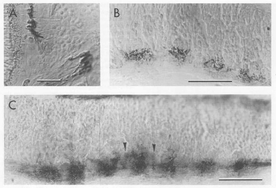
Fig. 4 Neuropil, Labeled by Znp Antibodies, First Develops in the Hindbrain Segment Centers
(A) Labeling by znp-1 is localized in the neuromuscular junction region of the extrinsic eye muscles, suggesting it recognizes a synaptic component. The other znp antibodies label muscle similarly and also show punctate staining at the bases of hair cells, where synapses have been identified (Metcalfe et al., 1985).
(B and C) Sagittal sections of the hindbrain at 48 hr. In (B), neuropil present in the segment centers is labeled in a coarse punctate fashion by znp-4. In (C), the znp-1 antibody labels the same region, but in a more delicate fashion. Longitudinal tracts are also labeled as well as a thin dorso-ventrally aligned region occasionally visible near the segment borders (arrowheads).
Scale bars: 25 μm.