
|
Fig. 6 Motor Nuclei, Labeled with Zn-5 at 48 h, Are in the Mi2 and Mi3 Segment Centers
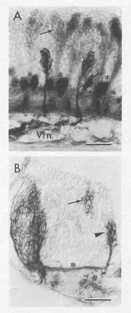
(A) The two motor roots exit the brain ventrally and join to form a single nerve that courses rostrally. The nerve (VI n.) is probably the abducens (cranial nerve VI), since trigeminal motor nuclei have been located in segments Ro2 and Ro3, and facial moto-nuclei have been located in segments Mi1 and Mi2 in 36 h embryos (T. Schilling and K. Hatta, unpublished data; also see Kimmel et al., 1985, for the locations of the same motor nuclei at 5 days). In chick embryos all of these nuclei are in the corresponding segments, including in particular the abducens motor nuclei in segments (R5 and R6; Lumsden and Keynes, 1989) that correspond to Mi2 and Mi3 in the fish. However, we have not directly verified the peripheral target of these labeled motoneurons. The large, darkly labeled bundles of commissural fibers (asterisks) mark the border regions of these segments, showing that the motor nuclei are in the centers. Another class of zn-5-positive neurons is located dorsally (arrow) in these segments, but not in more rostral ones. Their ventral running axons are in the border regions and may contribute to the ventral commissures.
(B) Transverse view of the left side of the hindbrain (dorsal up, lateral to the left) at the Mi2 segment, labeled with zn-5 at 48 h. The motor nucleus is located medially, near the ventricle (arrowhead). The dorsal goup of neurons (arrow) occupies a more medial position than the larger custer of neurons present in the sensory area to the left (the ones also shown in Figure 2) that give rise to the ventral commissure (asterisk).
Scale bars: 25 μm.