
- Title
-
Molecular heterogeneity among primary motoneurons and within myotomes revealed by the differential mRNA expression of novel islet-1 homologs in embryonic zebrafish
- Authors
- Tokumoto, M., Gong, Z.Y., Tsubokawa, T., Hew, C.L., Uyemura, K., Hotta, Y., and Okamoto, H.
- Source
- Full text @ Dev. Biol.
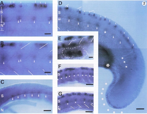
Developmental changes in the zfIsl-2 mRNA expression and comparison with the Isl-1 mRNA expression pattern. The segment borders of the somites are indicated by lines. S, spinal cord; N, notochord. Anterior, left, dorsal, up. (A, B) Lateral views of the same middle body trunk region of an 18-hr embryo focused to the spinal cord in A and to the somites in B. The dorsal and ventral zfIsl-2-positive cells are indicated by arrows and arrowheads, respectively. (C) A lateral view of the 15-hr embryo. By this stage, faint expression of zfIsl-2 mRNA had started in the ventral cells (arrowheads) located at the midsegments but not yet in the dorsal cells. The notochord was inadvertently displaced in this sample. (D) The caudal region of the same embryo as that shown in A and B at a lower magnification. In addition to the expression in the spinal cord. zfIsl-2 mRNA was observed along the apical ectoderm of the tail bud (dots) and in the region near the caudal end (asterisk) of the pronephric duct. The most caudal three segments are indicated by stars. No ventral cells caudal to this region expressed zfIsl-2 mRNA. (E) A close-up dorsolateral view of the pectoral fin bud region (surrounded by a dashed line) of a 30-hr embryo. zfIsl-2 mRNA was expressed in large round cells within the pectoral fin bud. The zfIsl-2 positive cells in the spinal cord are out of focus. (F, G) Isl-1 mRNA was expressed in large round cells within the pectoral fin bud. The zfIsl-2 positive cells in the spinal cord are out of focus. (F, G) Isl-1 mRNA expression in 15.5 (F) and 16 (G)-hr embryos for comparison. These are the lateral views of the same samples shown in our previous paper (Inoue et al., 1994). At 15.5 hr, Isl-1 mRNA was expressed in the ventromedial cells slightly anterior to the segment borders (indicated by white triangles) and weakly in the cells around the midsegments (indicated by white dots), in addition to the expression in Rohon-Beard cells. By 16 hr, Isl-1 mRNA expression in the ventral spinal cord was restricted only to the cells slightly anterior to the segment borders (indicated by white triangles). Bar, 25 μm (A, B, and E) and 50 μm (C, D, F, and G). |

zfIsl-3 mRNA expression in the body of a 19-hr embryo. (A) A photomontaged lateral view. Anterior, left; dorsal, up. (B) A cryostat cross section made at the rostrocaudal level indicated by the arrow in A. S, spinal cord; N, notochord. zfIsl-3 mRNA was expressed intensely in the ventral myotomes [brackets] and the Rohon-Beard neurons (RB) and weakly in the ventromedial regions (arrowheads) of the dorsal myotomes. Bar, 100 μm (A) and 25 μm (B). |
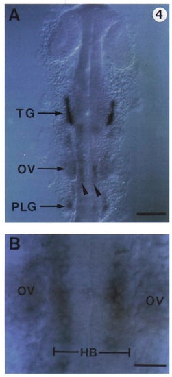
zfIsl-2 mRNA expression in the head of a 24-hr embryo. (A) A dorsal view. Anterior is up. Intense expression of zfIsl-2 mRNA was observed only in the trigeminal ganglia (TG). Weak expression was detected in the otic vesicles (OV), the posterior lateral line ganglia vesicles, showing the zfIsl-2-positive cells opposing the otic vesicles inside the hindbrain (HB). Bar, 100 μm (A) and 25 μm (B). |

zfIsl-3 mRNA expression in the head of a 24-hr embryo. (A) A lateral view. (B) A dorsal view. The zfIsl-3 mRNA expression was observed in the eyes (E) and the tectal region (T) of the midbrain as well as in the trigeminal ganglia (TG) and the ganglia anterior to the otic vehicle (AG). Otic vehicle, OV. Bars, 100 μm. |

Double labeling of embryos with the antibody (α-Tb) for acetylated α-tubulin and in situ hybridization to zfIsl-2 or zfIsl-3 mRNA. Anterior, left; dorsal, up; S, spinal cord; N, notochord. The somite borders are indicated by broken lines. (A, B) Photomontaged lateral views of the spinal cord at the middle body trunk level of two 24-hr embryos double-stained by in situ hybridization to zfIsl-2 mRNA and by α-Tb, α-Tb stains alost all axons and some cell bodies of neurons in the spinal cord by brown reaction products. A single or a pair of ventral zfIsl-2-positive cells (open triangles) at each midsegment had thin purple precipitates around the contours of the cell bodies. Their cell bodies were connected with motor nerve roots (open stars) by the axons (arrows) which extended straight ventrally inside the spinal cord. Axons (arrowheads) were observed to extend longitudinally from the dorsal zfIsl-2-positive cells and ultimately became confluent with the dorsal longitudinal fascicle (DLF) (indicated by brackets). The commissural neurons (denoted as C) were all negative for the zfIsl-2 mRNA expression. (C) Photomontaged lateral views of the spinal cord at the middle body trunk level of a 24-hr embryo double-stained by in situ hybridization to zfIsl-3 mRNA and by α-Tb. The dorsal zfIsl-3-positive cells extended the axons (arrowheads) longitudinally which ultimately became confluent with DLF (bracket). Bars, 25 μm. |

Double labeling of 22-hr embryos with a lineage tracer and in situ hybridization to zfIsl-2 mRNA. Anterior, left; dorsal, up; S, spinal cord. The somite borders are indicated by broken lines. The signals (indicated by dotted lines) for the zfIsl-2 mRNA expression in the ventral cells were detected as diffuse dark purple precipitates around the cell bodies. (A) A double-labeled ventral cell (asterisk) was characterized by intense brown cytoplasmic and axonal stainings and diffuse dark purple precipitates surrounding the cell body. It was identified as either CaP or VaP because of its large cell body located at the midsegment and the trajectory of the axon (arrows) that extended straight ventrally both inside and outside the spinal cord toward the ventral myotome. The neighboring small brown cells (arrowheads) were labeled only with biotin and had no dark purple precipitates around them. They were neuroepithelial cells since they had small cell bodies and no axons extending from them. A schematic drawing of the double-labeled cell is shown in the inset at the left bottom. The area stained by the signal for the zfIsl-2 mRNA expression is dotted. (B) A biotin-labeled commissural neuron (C) shown as a negative control for the zfIsl-2 mRNA expression. Bar, 25 μm. |
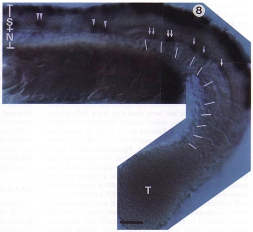
zfIsl-2 mRNA expression in the 24-hr spt mutant embryos. A photomontaged lateral view. Deficient cell migration during epiboly results in defective somites in the middle body trunk and the abnormal accumulation of cells at the end of the tail (T) in the spt mutant embryo. The embryo shown here has 10 apparently regular somites. The somite borders are indicated by lines. zfIsl-2 mRNA was not expressed in the ventral spinal cord caudal to the last 5 somites and expressed only in single or pairs of ventral cells (arrows) at the midsegments in the region surrounded by the anterior 5 of the 10 regular somites. In contrast, in the more anterior region where the somatic structure was severely disturbed, the zfIsl-2-positive ventral cells (arrowheads) significantly decreased in number per one segment length (0.5 in this mutant embryo compared to ∼1.5 in the normal embryos). S, spinal cord; N, notochord. Anterior, left; dorsal, up. Bar, 50 μm. |
Reprinted from Developmental Biology, 171, Tokumoto, M., Gong, Z.Y., Tsubokawa, T., Hew, C.L., Uyemura, K., Hotta, Y., and Okamoto, H., Molecular heterogeneity among primary motoneurons and within myotomes revealed by the differential mRNA expression of novel islet-1 homologs in embryonic zebrafish, 578-589, Copyright (1995) with permission from Elsevier. Full text @ Dev. Biol.