
|
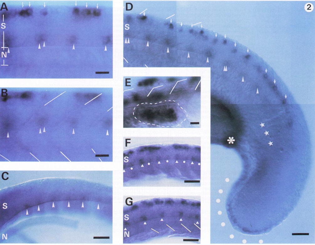
Fig. 2 Developmental changes in the zfIsl-2 mRNA expression and comparison with the Isl-1 mRNA expression pattern. The segment borders of the somites are indicated by lines. S, spinal cord; N, notochord. Anterior, left, dorsal, up. (A, B) Lateral views of the same middle body trunk region of an 18-hr embryo focused to the spinal cord in A and to the somites in B. The dorsal and ventral zfIsl-2-positive cells are indicated by arrows and arrowheads, respectively. (C) A lateral view of the 15-hr embryo. By this stage, faint expression of zfIsl-2 mRNA had started in the ventral cells (arrowheads) located at the midsegments but not yet in the dorsal cells. The notochord was inadvertently displaced in this sample. (D) The caudal region of the same embryo as that shown in A and B at a lower magnification. In addition to the expression in the spinal cord. zfIsl-2 mRNA was observed along the apical ectoderm of the tail bud (dots) and in the region near the caudal end (asterisk) of the pronephric duct. The most caudal three segments are indicated by stars. No ventral cells caudal to this region expressed zfIsl-2 mRNA. (E) A close-up dorsolateral view of the pectoral fin bud region (surrounded by a dashed line) of a 30-hr embryo. zfIsl-2 mRNA was expressed in large round cells within the pectoral fin bud. The zfIsl-2 positive cells in the spinal cord are out of focus. (F, G) Isl-1 mRNA was expressed in large round cells within the pectoral fin bud. The zfIsl-2 positive cells in the spinal cord are out of focus. (F, G) Isl-1 mRNA expression in 15.5 (F) and 16 (G)-hr embryos for comparison. These are the lateral views of the same samples shown in our previous paper (Inoue et al., 1994). At 15.5 hr, Isl-1 mRNA was expressed in the ventromedial cells slightly anterior to the segment borders (indicated by white triangles) and weakly in the cells around the midsegments (indicated by white dots), in addition to the expression in Rohon-Beard cells. By 16 hr, Isl-1 mRNA expression in the ventral spinal cord was restricted only to the cells slightly anterior to the segment borders (indicated by white triangles). Bar, 25 μm (A, B, and E) and 50 μm (C, D, F, and G).
Reprinted from Developmental Biology, 171, Tokumoto, M., Gong, Z.Y., Tsubokawa, T., Hew, C.L., Uyemura, K., Hotta, Y., and Okamoto, H., Molecular heterogeneity among primary motoneurons and within myotomes revealed by the differential mRNA expression of novel islet-1 homologs in embryonic zebrafish, 578-589, Copyright (1995) with permission from Elsevier. Full text @ Dev. Biol.