
|
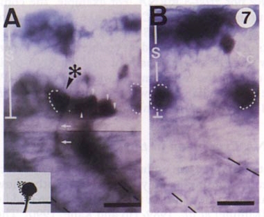
Fig. 7 Double labeling of 22-hr embryos with a lineage tracer and in situ hybridization to zfIsl-2 mRNA. Anterior, left; dorsal, up; S, spinal cord. The somite borders are indicated by broken lines. The signals (indicated by dotted lines) for the zfIsl-2 mRNA expression in the ventral cells were detected as diffuse dark purple precipitates around the cell bodies. (A) A double-labeled ventral cell (asterisk) was characterized by intense brown cytoplasmic and axonal stainings and diffuse dark purple precipitates surrounding the cell body. It was identified as either CaP or VaP because of its large cell body located at the midsegment and the trajectory of the axon (arrows) that extended straight ventrally both inside and outside the spinal cord toward the ventral myotome. The neighboring small brown cells (arrowheads) were labeled only with biotin and had no dark purple precipitates around them. They were neuroepithelial cells since they had small cell bodies and no axons extending from them. A schematic drawing of the double-labeled cell is shown in the inset at the left bottom. The area stained by the signal for the zfIsl-2 mRNA expression is dotted. (B) A biotin-labeled commissural neuron (C) shown as a negative control for the zfIsl-2 mRNA expression. Bar, 25 μm.
Reprinted from Developmental Biology, 171, Tokumoto, M., Gong, Z.Y., Tsubokawa, T., Hew, C.L., Uyemura, K., Hotta, Y., and Okamoto, H., Molecular heterogeneity among primary motoneurons and within myotomes revealed by the differential mRNA expression of novel islet-1 homologs in embryonic zebrafish, 578-589, Copyright (1995) with permission from Elsevier. Full text @ Dev. Biol.