
- Title
-
Connexin43 regulates joint location in zebrafish fins
- Authors
- Sims Jr, K., Eble, D.M., and Iovine, M.K.
- Source
- Full text @ Dev. Biol.
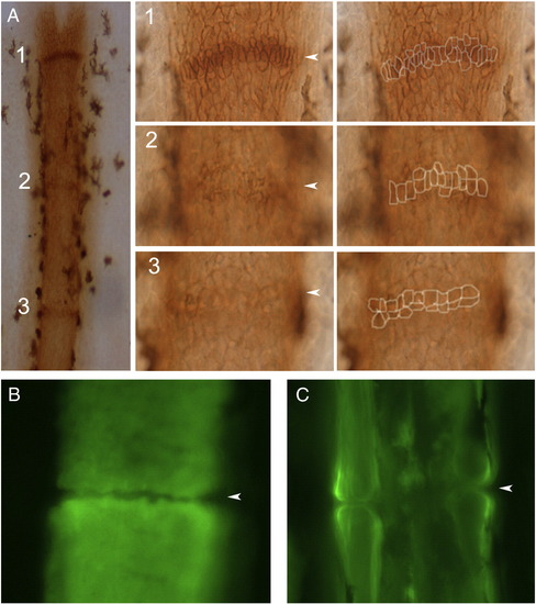
Joint formation in zebrafish fin rays. (A) Fins were stained with the osteoblast marker ZNS5, and detected using HRP (brown). At this level joints are detected as condensations of ZNS5-positive cells, which appear with differing maturities along the proximal–distal axis. Joint 1 is the most-distal joint and the least mature. At higher magnification distinct morphologies of ZNS5-positive cells can be observed (and outlines of the ZNS5-positive cells are shown to the right). (B) The morphology of the bone matrix in the newest joint is observed using calcein. (C) The morphology of the bone matrix in a fully mature joint is observed using calcein. Arrowheads point to joints. |
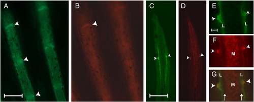
ZNS5 and Cx43 detect different populations of cells in the fin ray, and are both expressed in newly developing joints. (A) Whole mount staining of ZNS5. (B) Whole mount staining of Cx43. (C) Longitudinal cryosection showing lateral ZNS5 staining and detection of one joint (arrowhead). (D) Longitudinal cryosection showing mesenchymal Cx43 expression and detection of one joint (arrowhead). (E–G) Higher magnification of the joint observed in C and D shown for ZNS5, Cx43, and the overlap. Arrowheads point to joints; arrows indicate the location of bone matrix. L, lateral; M, medial. Scale bars for A–D, 100 μm. Scale bar for E–G, 10 μm. |

Cx43 localization during joint morphogenesis. A single image from a Z-stack collected by confocal microscopy is shown in the X–Y plane. The display on top represents a slice through the Z-stack along the horizontal green line (i.e., the X–Z plane). The blue line in this display indicates the location of the front image within the Z-stack. During elongation (A), Cx43 (arrows) is found surrounding the ZNS5 positive cells. During separation (B) and maturation (C), Cx43 appears polarized (brackets) toward the medial face of the ZNS5-positive cells. A group of five cells from the X–Z planes are represented by cartoons to the right of each image. Thick black bars over each X–Z plane identify the cells represented by cartoons at the right. Scale bar, 10 μm. |
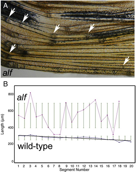
Segment length is irregular in alfdty86 fins. (A) Bright-field image of an alfdty86 fin. Arrows point to joints. (B) Segment length in wild-type and alfdty86 fins. Segment length was measured along the V + 3 fin ray in each of 10 fins. Segment length gradually decreases along the proximal–distal axis in wild-type fins, but appears random in alfdty86 fins. PHENOTYPE:
|

The cx43 mRNA is overexpressed in alfdty86 fins. (A, B) Whole mount in situ hybridization shows that the distal expression domain of cx43 is expanded in alfdty86 fins. (C, D) The expansion of the mesenchymal cx43 expression domain is also apparent following cryosectioning of stained fins. (E) The length of the cx43 expression domain was measured from the V + 3 fin ray in each of ten fins, and is statistically different between alfdty86 and wild-type fins. |

Targeted gene knockdown of cx43 restores joint formation in alfdty86 fins. (A) Segment length in alfdty86 fins following cx43 gene knockdown (alf KD) is similar to segment length following cx43 gene knockdown in wild-type fins (wt KD). Results of gene knockdown in both wild-type and alfdty86 are statistically different from wild-type fins treated with a non-targeting morpholino (p << 0.05). Wild-type and alfdty86 knockdown fins are not statistically different from one another. (B) Joints (arrows) are observed in each of three alfdty86 fin rays treated with cx43-MO2. |
Reprinted from Developmental Biology, 327(2), Sims Jr, K., Eble, D.M., and Iovine, M.K., Connexin43 regulates joint location in zebrafish fins, 410-418, Copyright (2009) with permission from Elsevier. Full text @ Dev. Biol.