
- Title
-
Endothelial signalling by the Notch ligand Delta-like 4 restricts angiogenesis
- Authors
- Leslie, J.D., Ariza-McNaughton, L., Bermange, A.L., McAdow, R., Johnson, S.L., and Lewis, J.
- Source
- Full text @ Development
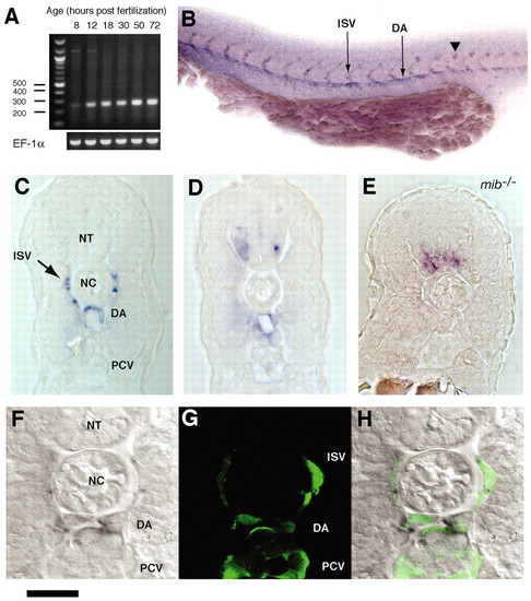
Expression of dll4 in wild-type and mutant zebrafish embryos. (A) RT-PCR analysis of whole homogenized embryos, showing weak dll4 expression as early as 8 hpf, becoming steadily stronger thereafter; EF-1α is a control. (B) Side view of a wild-type 25-hour-old embryo stained by in situ hybridization. dll4 is expressed in the dorsal aorta (DA) and intersegmental vessels (ISV), and in a subset of neurons in the neural tube (arrowhead). (C) Cross section through the trunk of a similar specimen showing dll4 expression in endothelial cells of the DA and ISVs but not in those of the posterior cardinal vein (PCV). (D) Similar section at a slightly different level relative to somite boundaries does not pass through any ISVs but reveals cells expressing dll4 in the ventrolateral neural tube (NT), as well as in the DA. (E) Cross section through the trunk of a 25-hour-old mib-mutant embryo. dll4 expression is increased and extended in the neural tube, but is undetectable in the endothelial cells. The expanded expression in the neural tube (and ear, for which data not shown) indicates that, in these tissues, dll4, like other delta genes, is regulated by lateral inhibition, so that its expression increases when Notch signalling fails. The loss of expression in the endothelial cells probably reflects their partial conversion to a venous character as a result of the failure of Notch signalling at an early stage (Lawson et al., 2001). The gross anatomy is also somewhat disturbed. (F-H) The neighbourhood of the notochord (NC) in a fli1:EGFP transgenic embryo, comparing dll4 expression (visible in the DA and ISVs by in situ hybridization; F,H) with the endothelium-specific expression of EGFP (green, visible by immunofluorescence in the DA, ISVs and PCV; G,H). Scale bar: 100 μm in B; 70 μm in C-E; 35 μm in F-H. EXPRESSION / LABELING:
PHENOTYPE:
|
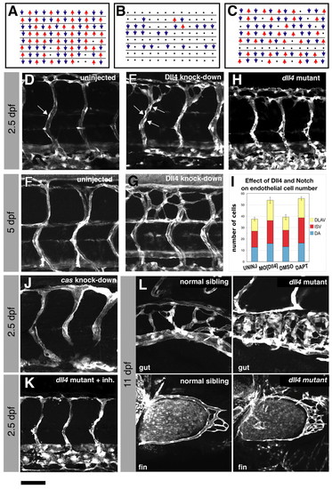
Effects of Dll4 knockdown on circulation and vascular pattern. (A) Blood flow in intersegmental vessels (ISVs) of wild-type living embryos at 3 dpf. Each line represents a set of ISVs on one side of one embryo, scored for direction of blood flow: upward arrows (red) denote flow from ventral to dorsal; downward arrows (blue), denote flow from dorsal to ventral; dots represent vessels carrying no flow. (B,C) Blood flow in intersegmental vessels of living embryos at 3 dpf injected with either 10 ng MO[Dll4] to knockdown Dll4 (B) or with 10 ng of a 5-base mismatch control morpholino (C). (D-G) fli1:EGFP embryos at 2.5 dpf (D,E) or 5 dpf (F,G) either uninjected (D,F) or injected with 10 ng MO[Dll4] (E,G). White blobs (arrows in D,E) are endothelial cell nuclei. (H) Similar region of a 2.5-dpf dll4-homozygous-mutant embryo (carrying the fli1:EGFP transgene). (I) Endothelial cell counts in the DAs, ISVs and DLAVs of embryos at 3 dpf. Embryos lacking Dll4 or treated with 100 μm DAPT have more cells than uninjected or DMSO-treated control siblings. Both effects are significant at the P=0.001 level (t-test; n≥6 specimens for each treatment; error bars represent s.e.m.). (J) fli1:EGFP embryo at 2.5 dpf injected with 10 ng of a morpholino targeted against casanova (sox32) to block circulation in the trunk. (K) fli1:EGFP-positive dll4-mutant embryo at 2.5 dpf treated from 48 hpf with 2 μM SU5416 to block Vegf signalling. (L) The gut and pectoral fin vasculature of an fli1:EGFP-positive dll4 mutant and a wild-type (normal) sibling embryo, both at 11 dpf. Scale bar: 50 μm in D-H,J,K; 40 μm in L. PHENOTYPE:
|

Effects of Dll4 knockdown on endothelial cell behaviour. (A) Schematic depiction of ISV and DLAV formation and the concomitant changes in endothelial cell morphology. (B,C) Leading cell of an ISV sprout at 30 hpf in a living uninjected embryo (B) or in a living embryo injected with 10 ng MO[Dll4] (C). (D,E) DLAV and dorsal ISVs at 2.5 dpf in a living uninjected embryo (D) or in a living embryo injected with 10 ng MO[Dll4] (E). (F-I) Similar region at 2.5 dpf of living embryos treated with 100 μm DAPT (G,I) or control embryos treated with DMSO (F,H). Treatment with DMSO or DAPT began at either 33 hpf (F,G) or at 48 hpf (H,I) and continued through the course of the experiment. Higher magnifications of D-I are shown in D'-I'. (J) DLAV and ISVs of a 3-dpf embryo injected with 7 ng of a morpholino targeted against Notch1b. (K,L) ISV sprouts at 30 hpf of a normal sibling (K) and a hsp70:Gal4;UAS:NICD embryo (L). Embryos in K and L were given a heat shock at 24 hpf. S, somite. Scale bar: 50 μm in B-I; 17 μm in D'-I'; 40 μm in J; 70 μm in K,L. PHENOTYPE:
|
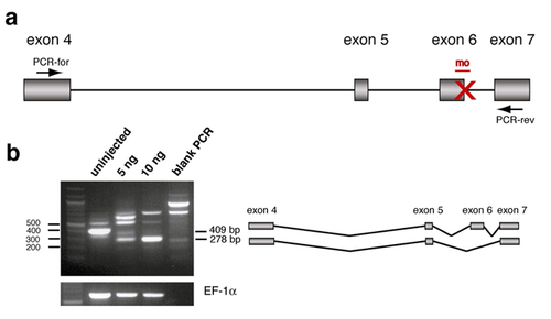
A splice-blocking morpholino targeted to dll4. (a) Schematic representation of the region spanning exon 4 through exon 7 of dll4. Target sites for MODll4 and the PCR primers used for RT-PCR are indicated. (b) RT-PCR analysis of dll4 splicing upon injection with 5 or 10 ng MODll4. In embryos injected with 10 ng MODll4, the normal transcript is undetectable and replaced by a predominant splice variant lacking exon 6 and with a frame-shift introducing a stop codon in exon 7. A total of 10 ng of the morpholino was used for all subsequent experiments. EF-1α was used as a loading control. The sequence for MODll4 is 5′-TAGGGTTTAGTCTTACCTTGGTCAC-3′. PCR was performed in two rounds using heminested primers. The first reaction was performed using the primer 5′-CATTGATTATTGAGGCCTGG-3′ (located 5′ of the region shown in a) and PCR-rev (5′-CTGTCACACTCTCGCACCTG-3′). A small amount of the resulting product was then amplified using PCR-for (5′-GTTTCGGCCACTACACCTGC-3′) and PCR-rev. |