
- Title
-
Notch inhibits Ptf1 function and acinar cell differentiation in developing mouse and zebrafish pancreas
- Authors
- Esni, F., Ghosh, B., Biankin, A.V., Lin, J.W., Albert, M.A., Yu, X., MacDonald, R.J., Civin, C.I., Real, F.X., Pack, M.A., Ball, D.W., and Leach, S.D.
- Source
- Full text @ Development
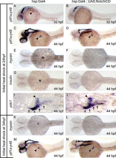
Notch pathway activation delays exocrine differentiation in developing zebrafish pancreas. Heat-shocked embryos expressing a hsp:Gal4 transgene alone or in combination with UAS:notch1aICD were assessed for initiation of ptf1a-p48 and trypsin expression, as well as expression of insulin and pdx1. Following heat shock at 24 hpf, ptf1a-p48 expression is delayed in 32 hpf hsp:Gal4;UAS:notch1aICD embryos (B) compared with hsp:Gal4 controls (A), but recovers by 34 hpf (data not shown). As assessed at 44 hpf, both trypsin (E,F) and insulin (G,H) expression are reduced in hsp:Gal4;UAS:notch1aICD embryos compared with hsp:Gal4 controls, even while ptf1a-p48 (C,D) and pdx1 (I,J) expression remain normal. Following delayed heat shock initiated at 34 hpf (after normal onset of ptf1a-p48 expression), ptf1a-p48 expression remains normal at all time points (M,N), whereas expression of trypsin is delayed (K,L). Arrowheads in A,C,D,M,N indicate endodermal domain of ptf1a-p48 expression, distinct from expression in developing hindbrain. Arrowheads in I and J indicate pdx1-positive principal islet; arrows indicate adjacent pdx1-positive intestine. |
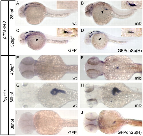
Defects in Notch pathway activation result in acceleration of exocrine differentiation in developing zebrafish pancreas. (A-D) Mindbomb (mib) mutant embryos (B) and embryos injected with RNA encoding a dominant-negative Suppressor of Hairless DNA binding mutant [GFPdnSu(H)] (D) show either accelerated (mib) or normal [GFPdnSu(H)] onset of ptf1a-p48 expression compared with clutchmate controls (A,C). Arrows indicate endodermal domain of ptf1a-p48 expression, distinct from expression in developing hindbrain. Insets in A-D show magnified view of endodermal ptf1a-p48 expression domain. (E-J) mindbomb (mib) mutant embryos (F,H) and embryos expressing GFPdnSu(H) (J) show accelerated acinar cell differentiation compared with clutchmate controls (E,G,I), marked by early onset of trypsin expression. Note normal absence of trypsin expression in control embryos at 40 hpf (E,I), but accelerated onset of trypsin expression in mibta52/ta52 and GFPdnSu(H)-injected embryos (F,J). At 80 hpf, the size and contour of established trypsin-positive exocrine parenchyma is also altered in mibta52/ta52 embryos compared with wild-type clutchmates (G,H). Wt indicates wild-type clutchmates arising from mibta52/wt x mibta52/wt cross. GFP indicates clutchmate control embryos injected with RNA encoding GFP alone. EXPRESSION / LABELING:
|