
- Title
-
Expression of the zebrafish genome during embryogenesis (NIH R01 RR15402)
- Authors
- Thisse, B., Pflumio, S., F�rthauer, M., Loppin, B., Heyer, V., Degrave, A., Woehl, R., Lux, A., Steffan, T., Charbonnier, X.Q. and Thisse, C.
- Source
- Submitted By
- F�rthauer, Maximilian, Thisse, Bernard, Thisse, Christine (Citing this work)
- Protocol
- Thisse in situ hybridization protocol
- Probe
- cb110 Quality:
- Supplier


Fig. 1 Expression of fgf8 starts soon after the midblatula transition in cell of the dorsal margin. Expression then spread from this dorsal position to encompass the whole margin at late blastula stage. This marginal expression is observed in deep cell layer as well as in EVL but not in YSL. EXPRESSION / LABELING:
|


Fig. 2 At the onset of gastrulation the expression of Fgf8 disappears progressively from the ventral margin and increases in dorsal marginal cells establishing a dorsal to ventral gradient of expression. At 60% epiboly a weak expression is still observed in EVL cells that are now located vegetaly to the marginal cells. Dorsally a strong expression is observed in presumptive paraxial mesodermal cell and in the deepest cells of the epiblast in direct contact with the involuted mesoderm. At mid-gastrulation, in addition to the marginal EVL and the embryo margin a novel site of expression appears in the presumptive hindbrain and the underlying cephalic paraxial mesoderm. A strong expression is also observed in ventral neurectoderm (presumptive floor plate). Expression of Fgf8 increases in presumptive hindbrain and disappears from paraxial cephalic mesoderm at late gastrula. Near the margin expression is strong in presumptive neurectodermis as well as in dorsal marginal cells (that give rise to axial structures). EXPRESSION / LABELING:
|


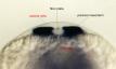


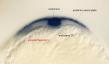


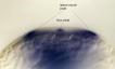


Fig. 3 The strong stripe in presumptive hindbrain progressively restrict to rhombomere 4 and the ventral part of rhombomere 2. Heart primordia appear labeled by the beginning of somitogenesis. At the 2/3 somite stage, the floor plate, posterior and lateral neural plate appear labeled as well as the caudal most axial tissues. In cross sections, cells located underneath the posterior notochord (endoderm?) are labeled. In hindbrain region, expression stay in the whole rhombomere 4 but is restricted to the ventral most part of rhombomere 2. Around the 3/4 somite stage, anterior expression start to encompass the midbrain-hindbrain boundary. In the paraxial mesoderm, Fgf8 expression is observed in newly formed somites. Anteriorly, after the 2 somite stage, strong labelling is observed in the telencephalon. At the 5 somite stage, expression is strong in the telencephalon, the midbrain-hindbrain boundary, in posterior rhombomere 2 (or anterior part of the cerebellum (?)), ventral part of rhombomere 2. Expression is observed in dorsal and ventral but not the central part of Rhombomere 4. Expression is still observed in heart primordia. In truncal region Fgf8 expression is observed in paraxial somitic mesoderm but not in the adaxial cells. Staining is maintained in the floor plate. By the 8 somite stage, expression has disappeared from the rhombencephalon but persists at the midbrain-hindbrain boundary. EXPRESSION / LABELING:
|


Fig. 4 At the 12 somite stage, expression is observed at the midbrain-hindbrain boundary, somites, posterior spinal cord, posterior floor plate, caudal tip of the notochord and cells ventral to it (endoderm?). Anteriorly, a strong staining is observed in the telencephalon, in head epidermis and in the optic stalk. A weak expression is observed in the ventral posterior diencephalon. At the 14 somite, expression appear in the retina as well as in the dorsal diencephalon. In the somites Fgf8 expression disappears from the posterior part and is restricted to the anterior cells lining the somitic furrow. EXPRESSION / LABELING:
|


Fig. 5 At 20 somite stage, expression increases in the proximal part of retina and the ventral diencephalon. Strong staining is observed in telencephalon, midbrain-hindbrain boundary, optic stalk and the dorsal diencephalon. Moreover, staining in now observed in ventral anterior mesencephalon. In the ear, fgf8 is expressed in the anterior most as well as the dorsal-posterior cells of the otic capsule. Expression is now observed in hyoid arch. Posteriorly, staining is observed in caudal somites as well as in dorsal and caudal fin fold. At 30 hrs expression is also observed in the nose. EXPRESSION / LABELING:
|


Fig. 6 At 36 hrs, expression is observed in the ganglion cell layer of the retina, the adenohypophysis, and the hyoid arch. Expression has disappeared from the posterior otic vesicle and the somites. An increase of expression is observed in median fin fold |


Fig. 7 At 48hrs, expression territories are unchanged compared to the expression at 36 hrs. |