
|
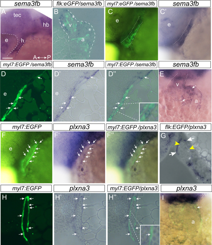
Fig. 1
sema3fb is expressed by embryonic cardiomyocytes. A-E Whole mount (A,C,C’,E) and horizontal (B) and sagittal (D-D’’) sections of sema3fb ISH on 28 hpf (A,C,D), 36 hpf (B) and 48 hpf (E) zebrafish hearts. sema3fb is expressed through the heart chamber, in GFP positive cardiomyocytes in a Tg(myl7:EGFP) heart (C–C’,D-D’’) and surrounding (asterisks) GFP positive endocardial cells in a Tg(flk:EGFP) heart (B). Blends between the GFP and ISH signals (B,C,D’’). Inset (D’’) shows a sema3fb-expressing cardiomyocyte. F-I Whole mount (F-F’’,I) and sagittal (G-H) sections of plxna3 ISH of 28 hpf (F-F’’,H–H’’), 36 hpf (G) and 48 hpf (I) zebrafish hearts. plxna3 mRNA is present in GFP positive cardiomyocytes (arrows) in a Tg(myl7:EGFP) heart (F-F’,H–H’’) that surround the GFP positive endocardial cells (yellow arrowheads) in a Tg(flk:EGFP) heart (G). Blend between the GFP and ISH signals (F’’,G,H’’,I). A, anterior; a, atrium; e, eye; h, heart; hb, hindbrain; P, posterior; Tec, optic tectum; v, ventricle. Scale bar: 50 µm (A,C-D,F–H), 25 µm (B,E,I)