Figure Caption
Figure 1
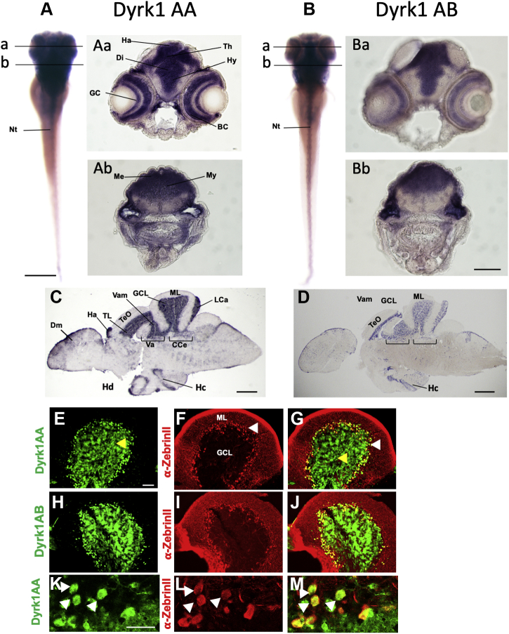
Ubiquitous expression of dyrk1aa and dyrk1ab in the larval nervous system becomes confined to specific structures in the adult brain. RNA in situ hybridization in zebrafish larvae and adult brain sections with dyrk1aa (A, Aa, Ab, C, and E–G) and dyrk1ab (B, Ba, Bb, D, and H–J) specific probes and respective sense controls are shown in Fig. S1. dyrk1aa and dyrk1ab both are expressed throughout the central nervous system of 5 dpf zebrafish larvae (A and B). Transverse sections from different planes with strong expression for both paralogs in the entire gray matter (Aa, Ab, Ba, and Bb). Chromogenic mRNA in situ of adult brain sagittal sections for dyrk1aa (C) and dyrk1ab (D) with intense staining in the cerebellar granule cell layer and the torus longitudinalis. Fluorescent dyrk1aa (E–G and K–M) and dyrk1ab (H–J) in situ with subsequent ZebrinII immunohistochemistry confirms expression in the granule cell layer below the Purkinje cell layer but also reveals expression in Purkinje cells, which was further confirmed by confocal microscopy at cellular resolution in which dyrk1aa expression (K) colocalizes with ZebrinII expression in individual Purkinje cells (L and M, white arrows). dyrk1aa (E and K), and dyrk1ab (H) fluorescent mRNA in situ (green) followed by anti-ZebrinII antibody staining (F, I, and L; red) with overlay for dyrk1aa (G and M) and dyrk1ab (J). Black lines in panels A and B indicate the plane of sections shown in panels Aa, Ab, Ba, and Bb. Yellow and white arrowheads indicate granule cells (GCs) and Purkinje cells (PCs), respectively. The scale bars indicate 200 μm (A and B), 100 μm (Aa, Ab, Ba, Bb, and E–J), 500 μm (C and D), 100 μm (E–J), and 20 μm (K–M). Bc, bipolar cells; CCe, corpus cerebelli; Di, diencephalon; Dm, medial zone of the dorsal telencephalic area; GCL: granule cell layer; Ha, habenula; Hc, central zone of the dorsal telencephalic area; Hd, dorsal zone of the periventricular hypothalamus; Hy, hypothalamus; La, lobus caudalis; Me, metencephalon; ML, molecular layer; My, myelencephalon; PCL, Purkinje cell layer; TeO, tectum opticum; Th, thalamus; TL, torus longitudinalis; Va, valvula cerebelli; Vam, medial division of valvula cerebelli.
Acknowledgments
This image is the copyrighted work of the attributed author or publisher, and
ZFIN has permission only to display this image to its users.
Additional permissions should be obtained from the applicable author or publisher of the image.
Full text @ J. Biol. Chem.