
Image
|
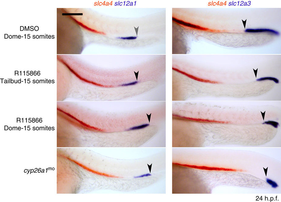
Figure Caption
Fig. 5
Knock down of Cyp26a1 anteriorizes the kidney.
Whole-mount double in situ hybridization analysis at 24 hours post fertilisation (h.p.f.) for nephron segment markers slc4a4 (orange), slc12a1 and slc12a3 (purple) in embryos injected with cyp26a1 morpholino or treated with R115866 or DMSO (vehicle control). Embryos are shown as lateral views with anterior to the left. Arrowhead indicates junction between the DE and DL segments. Scale bar, 100 µm.
Figure Data
Acknowledgments
This image is the copyrighted work of the attributed author or publisher, and
ZFIN has permission only to display this image to its users.
Additional permissions should be obtained from the applicable author or publisher of the image.
Full text @ Nat. Commun.